
Effects of Glutamine and Glycine Supplementation on Invitro Vitrification of Immature bovine oocytes
Abstract
The present study was designed to investigate the effects of supplementation of the maturation media with glutamine and glycine on the in vitro maturation of immature oocytes. Different concentrations of glutamine and glycine (0.5, 1, 2 mM) were used in the current study. The oocytes’ morphology, viability, cleavage, and blastocyst rates were the dependent variables in the present study (p<0.05). The highest morphology score was reported when using when1 mM glutamine and glycine compared to control (p<0.05). Addition of 0.5Mm glutamine and glycine did not significantly differ from the control group. Moreover, addition of 2Mm glutamine and glycine significantly decreased normal morphology % when compared to the control group. The highest viability rate was reported when using 1mM of glutamine and glycine (p<0.05) which is significantly different than control and 0.5mM. Addition of 2mM significantly decreased viability when compared to the control group. The highest cleavage rate was reported when using 1mM glutamine and glycine which was significantly increased when compared to the control group (p<0.05). Addition of 0.5mM glutamine and glycine significantly increased cleavage rate when compared to the control group (p<0.05). Addition of 2mM glutamine and glycine significantly decreased cleavage rate when compared to the control group. The highest blastocyst rate was reported when using 1mM glutamine and glycine which significantly increased blastocyst formation rate when compared to control. Addition of 0.5mM glutamine and glycine significantly increased this parameter when compared to the control group but addition of 2mM result didn’t differ from control (p>0.05). The highest morphology score, viability rate, cleavage rate and blastocyst formation rate were reported when using 1mM glutamine and glycine.
Article Information
- Received
- Accepted
- Published
Academic Editor: John Okyere, CrossGen Limited
Checked for plagiarism: Yes
Copyright © 2024 Mona M Trabih, et al.
 This is an open-access article distributed under the terms of the Creative Commons Attribution License, which permits unrestricted use, distribution, and reproduction in any medium, provided the original author and source are credited.
Corresponding author: Mohammed A Elmetwally, DMV, Department of Theriogenology, Mansoura University, Mansoura, 35516, Egypt —
Competing Interests
The authors have declared that no competing interests exist.
Funding
No specific funding statement was provided by the authors.
Data Availability
No data-availability statement was provided by the authors.
Citation:
Introduction
Glutamine and glycine are basic amino acids (AA) present in the physiological fluids of all living things. They do this by activating cellular target of rapamycin (MTOR) signaling. This increases protein synthesis, reduces protein production, and 1 promotes the development of brown adipose tissue during ruminant mammal conception 2, 3, 4.
When glutamine and glycine are present in the ruminant body, they have many important effects on nitrogen metabolism, blood circulation, nutrient utilization and ruminant health 1, 5 Rumen bacteria produce this amino acid. In addition, thanks to the process of manufacturing L-citrulline (Cit) from these three elements, L-glutamine, L-glutamate and L-proline are also produced in the enterocytes of young and adult ruminants 6, 7. In pre-weaning ruminants, most of the Cit initially produced by enterocytes is used in the form of glutamine and glycine. In ruminants after weaning, small intestine-derived Cit is mainly converted to glutamine and glycine in the kidneys, but also in endothelial cells, macrophages, and other cells 3, 7. According to the normal diet, the synthesis of glutamine and glycine accounted for 66% and 68%, respectively, of the total glutamine and glycine requirements for the diets of non-pregnant women and late-term pregnant women, which contain less than 12% protein 9. Creatine production requires 40% and 36% of the Arg used by non-pregnant women and pregnant women in late pregnancy, respectively. Glutamine and glycine are not considered prohibited in the diet of ruminants after weaning, during gestation or during pregnancy, because it is believed that these animals can produce enough glutamine and glycine to meet their nutritional and physical needs. Because there is not enough information on the nutrition and metabolism of glutamine and glycine, ruminants do not get the best milk, reproduction and growth from their bodies 7. There is now ample evidence showing that rumen-protected glutamine and glycine can help improve all of these productions without affecting metabolism or health. For example, 0.25-0.5% dry food should be added to the food. Since bacteria in the rumen do not break down extracellular Cit because it does not enter the rumen, Cit can be used as a source of glutamine and glycine for ruminants such as cattle, cows, sheep and goats without encapsulation 8. Rumen - Glutamine and glycine or unencapsulated Cit are abundant because ruminants need them not only to help them live, grow, produce milk, produce and feed more, but also to keep them healthy and happy 8, 10. In vitro embryo production in cattle has received much attention. Indeed, it can be used for research and business purposes. The transfer of embryos into the cow takes a long time. This is because buffaloes do not have a good super-ovulatory response. Aspiration and excision are the two most common methods for obtaining oocytes from slaughtered cow ovaries 11. Fertility services such as ovarian hyper-stimulation (superovulation), immature oocyte collection, in vitro maturation (IVM), artificial insemination (AI), IVF and embryo transfer (ET) are available and applied to Bovidae spp. This increases the number of offspring of selected females and shortens the generation 11. Ovaries from a slaughterhouse are the cheapest source of eggs. This means that embryos can be produced in large quantities and at low cost. To elucidate the role of protein recruitment in oocyte maturation, we investigated the effects of various proteins on oocyte maturation and subsequent embryo development following IVF.
Material and Methods
The study was carried out at Animal Reproduction Research Institute - Giza, 12556 Al Ahram -Giza, 12111 Cairo – Egypt during the period from December 2022 to April 2023. The chemicals in this study will be purchased from Sigma Chemical Co (St. Louis, MO, USA). All experimental protocols have been approved (M\34) by the Committee for Research Ethics at the Faculty of Veterinary Medicine Mansoura University, Egypt.
Biological materials
a- Ovaries
Ovaries from apparently normal reproductive organs of cows of unknown age and breeding history were collected within 30 minutes after slaughter and evisceration of animals at the private ELsharkawy abattoir. The ovaries were kept in a thermos flask containing warm normal saline, then transported to the lab within 1-2 h after slaughtering 12. The ovaries were further washed as soon as we arrived at the lab in warm normal saline to remove the blood and debris and then kept in a water bath at 37°C during oocyte collection according to the previous literature 3, 13, 14.
b- Oocytes
Immature oocytes were collected by aspiration of medium-sized (4- 8 mm ) ovarian follicles using an 18-gauge needle attached to a disposable 10 ml syringe according to the previous studies 13, 15. Evenly granulated oocytes surrounded with one layer of cells were selected for further experimental procedures. The selected immature oocytes were firstly washed three times in sterile (PBS) phosphate buffer saline solution.
c- Semen
Frozen cow semen was obtained from Animal Reproduction Research Institute.
Chemical material
The chemicals used in this study were purchased from Sigma Chemical Co (St. Louis, MO, USA).
Preparation of basic media
Preparation of media and stock solution requires a sterile technique with accurate and careful weighing of components. The use of a laminar flow cabinet is indispensable to avoid any contamination that may alter and spoil the prepared media. An analytical balance with readability of at least 10 μg and accuracy of ± 0.1μg was used. All media were filtered using a 0.2 μm (Millipore, USA) syringe filter and incubated for at least 2 h in a humidified atmosphere, 5% CO2 at 38.5° C before culturing the oocytes and spermatozoa.
Basic medium (BM)
The basic medium used for all vitrification and warming solutions in this study was tissue culture media 199 (TCM 199) supplemented with 20%v/v fetal calf serum.
Vitrification solutions
Basic medium plus 50% v/v of final CPAs concentration was used as an equilibration solution (ES), while vitrification solutions (VS) were made of basic media containing the final CPAs concentration with or without 0.5 M sucrose.
Warming solutions (WS)
Three warming solutions were prepared from basic medium plus 0.5M sucrose .
In vitro maturation medium
In vitro fertilization medium
Recovery and classification of immature oocytes
In the lab, ovaries were washed in fresh, sterile physiological saline to further remove any contaminants. Then the ovaries were dried with Material and methods 31 sterile paper towels and by the use of an 18-gauge needle attached to a 10 ml syringe follicles (4 to 8 mm ) were aspirated and pooled in a 15 ml conical tube. The tubes are left 10-15 minutes after the end of aspiration to make their content sediment. The large follicles should not be aspirated because it causes jelly formation in the aspiration which affects oocytes recovery and searching. After sedimentation, about 5ml of sediment was aspirated and placed in a 10 cm diameter polystyrene sterile petri dish 3, 15.
As much as possible recovery of oocytes must occur in sterile conditions at room temperature. The immature oocytes were recovered and selected by using a stereomicroscope (The Russian M3 Stereomicroscope; magnification power 25 X) and picking to oocytes in a sterile automatic pipette and transferred into another dish containing fresh pre-warmed washing media 16
Vitrification of immature oocytes
Equilibration and Vitrification solutions were prepared by using TCM 199 medium supplemented with 10% FCS as a basic medium. Five immature oocytes were placed in 50μl drops of basic medium for one minute then transferred to equilibrated solution (100μl drops of basic media with added 10% serum+ 10% EG+10% DMSO) for 15 minutes at room temperature.
Then equilibrated oocytes were transferred to vitrification solution VS (100μl drops of basic media with added 10% serum + 20%EG + 20% DMSO). During vitrification glutamine and glycine were added at different concentrations (control group;0.5,1,2mM glutamine and glycine) for 30 sec. 3, 13.
Oocytes were immediately loaded in a closed straw. five to ten oocytes were immediately loaded in 0.25-ml mini-straws in the middle column of the vitrifying solution separated by air bubbles and sealed with polyvinyl alcohol ( PVA) powder. Then the straws were pre-cooled by keeping them in LN2 vapor at a height of about 5 cm from LN2 to avoid cracking of straws for one min; following this they were dipped vertically in LN2.
Warming of vitrified cow oocytes
After a period of storage (1 month), the vitrified oocytes in a straw were thawed in a water bath at 37°C for one min and the content of each straw was expelled into a tissue culture dish containing 0.5 ML sucrose solution and held for 3 min for stepwise rehydration. Finally, the immature oocytes were incubated in basic medium (TCM199) for 3min at room temperature.
Evaluation of vitrified/warmed immature oocytes
quality Oocyte recovery rate: The number of oocytes counted after the end of rehydration, in relation to the total of vitrified oocytes 17.
a-Evaluation of oocytes’ morphology Vitrified /warmed oocytes
were examined under a stereo microscope for evaluation of normal and abnormal oocytes according to 3. The oocytes with spherical and symmetrical shape and no signs of lyses/degeneration were considered normal whereas oocytes with ruptured zona pellucida, fragmented cytoplasm, or degenerative signs were classified as abnormal and discarded.
b-Evaluation of viability using the trypan blue exclusion test
Trypan blue stain is a useful and quick method to assess the initial quality and viability of oocytes. Trypan blue solutions (0.05%) were prepared by dissolving trypan blue in phosphate buffer saline (PBS; pH 7.0). Oocyte staining was performed at room temperature for 2 min 18. The exclusion test using trypan blue stain provides an assessment of cell membrane integrity as those cells with damaged or non-intact cell membranes permit the passage of the trypan blue toward the nucleus of an oocyte. Oocytes were categorized on the basis of the degree of dye exclusion. Unstained oocytes were classified as live and fully stained oocytes as dead 19.
In vitro maturation of the vitrified-warmed oocytes
Good quality immature oocytes were rinsed three times in maturation media TCM and each 10- 15 oocytes transferred to a 100 μl drop of maturation media under mineral oil and put in an incubator for 20-22 hours at 38.5° C in 5% of CO2 in air with maximum humidity.
After warming, viable oocytes were washed twice in maturation medium: TCM 199 supplemented with 10% FCS, 10 μg/mL LH, 5 μg/mL FSH and 50 μg/mL gentamycin sulfate. Groups of 20−25 oocytes were then cultured in pre-warmed maturation medium under mineral oil for 22 h at 38.5ºC, in 5% CO2 in air.
Assessment of maturation rate
Maturation rate was assessed under a stereomicroscope by expansion of cumulus cell around the oocyte. An oocyte with complete or moderate cumulus expansion and also the oocyte with slight or no cumulus cell expansion but has the first PB extruded in the perivitelline space were considered matured 18, 20.
Semen preparation and oocyte fertilization
Firstly, a fertilization dish was prepared by adding 50 μl modified Tyriod's Albumin Lactate Pyruvate (TALP) media drops in a sterile disposable petri dish then covered with mineral oil and incubated at 38.5 ◦C in 5% CO2 in air with maximum humidity.
Mature oocytes were partially denuded from the surrounding cumulus cell to allow easy penetration of the sperm by repeated gentle pipetting and washed three times in TALP media then added to the fertilization drops (10 oocyte/drop).
Spermatozoa were capacitated in vitro using TALP medium. Three straws of frozen semen with known sperm cell concentration of about (2×10⁶ sperm cell/ml) were thawed for one minute in a 37°C water bath. Immediately after thawing, by swim up technique the most motile spermatozoa were separated in sperm–TALP medium containing 6 mg/ml BSA, for one hour 21. Then the uppermost layer of the medium containing the most motile spermatozoa was collected by using a plastic Pasteur pipette and transferred into a small test tube containing 3 ml of SP-TALP medium.
The freezing media of the sperm is washed out by centrifugation at 2000 round per minute (rpm) for 5 minutes to form a sperm pellet; the supernatant is discarded and one ml of TALP is added and mixed with pellet then centrifuged and the supernatant is discarded again to completely wash out the freezing media.
One ml TALP medium containing 10 mg /ml heparin for in vitro capacitation of sperm is added and mixed with the sperm pellet and incubated at 38.5 ◦C in 5 % of CO2 and air with maximum humidity in an incubator for 10 minutes then checked for motility. If motility is acceptable, 20 μl of semen was co-incubated with each fertilization drop for 18 hours 3, 15.
In vitro Embryo development
Firstly, the culture dish was prepared by adding 50μl culture media drops in a sterile disposable petri dish then covered with mineral oil and incubated at 38.5 ◦C in 5% of CO2 in a maximum humidity incubator. The oocytes were freed from loosely bound spermatozoa and remaining attached cumulus cells by gentle pipetting.
All the culture media performed in an atmosphere of CO2 in air with maximum humidity at 39°C. The culture medium was replaced every 48 h with a fresh medium until day 5-7 post-insemination to prevent toxic accumulation of ammonium as a result of amino acid degradation and oocytes that had not cleaved must be removed from the culture media, leaving only those that undergo cleavage. During the observation period, gentle shaking of the culture dish was done to allow a uniform environment among fertilized oocytes.
Assessment of fertilization and embryonic development
At 18 h following insemination, the presumptive zygotes were denuded completely from surrounding cumulus cells and mounted on slides with coverslips and then fixed with acetic acid/ethanol (1:3) solution for at least 24 h. The presumptive zygotes were stained with 1% orcein dissolved in 45% acetic acid solution and examined for evidence of fertilization. Penetration of the sperm was identified by observing decondensed sperm heads or male pronuclei with their accompanying sperm tails in the cytoplasm.
Oocytes with two pronuclei and a clear second PB were considered normally fertilized. The percentage of cleaved oocytes was assessed at 48 h after insemination. On day 5 and day 7 following insemination, the embryos were observed under a microscope to compare the percentages of embryonic development to morula in each group respectively.
Experimental design
Experiment 1. Effect of Glutamine and Glycine addition during vitrification on morphology of vitrified/warmed cow oocytes:
The immature oocytes were divided into 4 groups according to the concentration of glutamine and glycine added to Vitrification solution to assess its effect on oocytes’ morphology as follows:
Group 1: vitrified oocytes without glutamine and glycine addition as control.
Group 2: vitrified oocytes supplemented with 0.5mM/ml glutamine and glycine.
Group 3: vitrified oocytes supplemented with 1mM/ml glutamine and glycine.
Group 4: vitrified oocytes supplemented with 2mM/ml glutamine and glycine.
Experiment 2. Effect of glutamine and glycine addition during Vitrification on viability of vitrified/warmed cow oocytes:
The immature oocytes were divided into 4 groups according to the concentration of glutamine and glycine added to Vitrification solution to assess its effect on oocytes’ viability as follows:
Group 1: vitrified oocytes without glutamine and glycine addition as control.
Group 2: vitrified oocytes supplemented with 0.5mM/ml glutamine and glycine.
Group 3: vitrified oocytes supplemented with 1mM/ml glutamine and glycine.
Group 4: vitrified oocytes supplemented with 2mM/ml glutamine and glycine.
Experiment 3. Effect of glutamine and glycine addition during vitrification on cleavage of vitrified/warmed cow oocytes:
The immature oocytes were divided into 4 groups according to the concentration of glutamine and glycine added to vitrification solution to assess its effect on oocytes’ cleavage as follows:
Group 1: vitrified oocytes without glutamine and glycine addition as control.
Group 2: vitrified oocytes supplemented with 0.5mM/ml glutamine and glycine.
Group 3: vitrified oocytes supplemented with 1mM/ml glutamine and glycine.
Group 4: vitrified oocytes supplemented with 2mM/ml glutamine and glycine.
Experiment 4. Effect of glutamine and glycine addition during vitrification on blastocysts of vitrified/warmed cow oocytes:
The immature oocytes were divided into 4 groups according to the concentration of glutamine and glycine added to vitrification solution to assess its effect on oocytes blastocysts as follows:
Group 1: vitrified oocytes without glutamine and glycine addition as control.
Group 2: vitrified oocytes supplemented with 0.5mM/ml glutamine and glycine.
Group 3: vitrified oocytes supplemented with 1mM/ml glutamine and glycine.
Group 4: vitrified oocytes supplemented with 2mM/ml glutamine and glycine.
Statistical analysis
Each experiment was at least three times replicated. According to Hussein et al. 2021 and 2019 22, 23. The normality of quantitative parameters was tested using normal probability plots and the Kolmogorov-Smirnov test created with SAS's UNIVARIATE technique. All experimental results are shown as mean SEM. The recovery rate, cleavage and maturation rate, and blastocyst rate are all represented as percentages. SAS® (version 9.2, SAS Institute, Cary, NC, USA) will be used for statistical analyses. Differences will be judged significant when they reach (P ≤ 0.05).
Results
Effect of Glutamine and Glycine addition during vitrification on morphology of vitrified/warmed cow oocytes:
The normal morphology score of cow oocytes after vitrification/warming by using different treatments of glutamine and glycine was determined (Table 1, Figure 1). The highest morphology score was reported when using 1 mM glutamine and glycine compared to control (86.27 ±3.42 vs 77.23 ± 4.19 respectively). Addition of 0.5Mm glutamine and glycine did not significantly differ from the control group (78.79± 4.13 vs 77.23 ± 4.19). Moreover, addition of 2Mm glutamine and glycine significantly decreased normal morphology % when compared to the control group (70.00 ±4.60 vs 77.23 ± 4.19).
Table 1. Effect of Glutamine and Glycine addition during vitrification on morphology of vitrified/warmed cow oocytes| Group | No. of oocytes | Normal morphology score (%) |
| Group 1 | 101 | 77.23 ± 4.19ab |
| control | ||
| Group2 | 99 | 78.79± 4.13 ab |
| 0.5mM | ||
| Group3 | 102 | 86.27 ±3.42 a |
| 1mM | ||
| Group4 | 100 | 70.00 ±4.60 b |
| 2mM |
Figure 1. Effect of glutamine and glycine addition during vitrification on morphology of vitrified/warmed cow oocytes. The data is presented as mean ± SD. Different letters a, b, & c indicate a significant difference (P≤0.05) between treatment groups.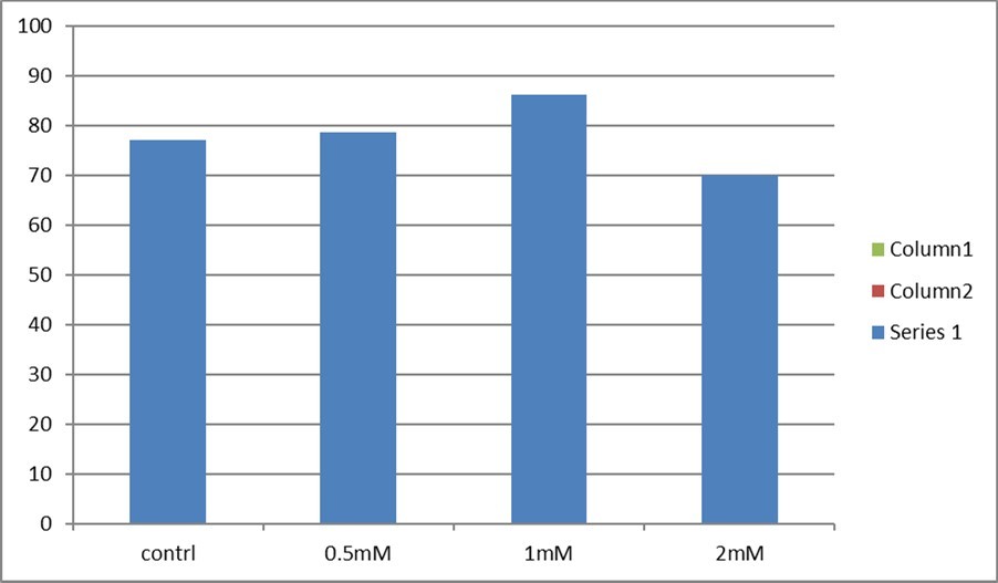
Download figure
Effect of glutamine and glycine addition during Vitrification on viability of vitrified/warmed cow oocytes:
The viability rate of bovine oocytes after Vitrification by using different treatments of glutamine and glycine was determined (Table 2, Figure 2). The highest viability rate was reported when using 1mM of glutamine and glycine (86.27 ±3.42a) which is significantly different than control and 0.5mM (78.79± 4.13 ab and 77.23 ± 4.19ab). Addition of 2mM significantly decreased viability when compared to the control group (70.00 ±4.60b vs 77.23 ± 4.19ab).
Table 2. Effect of glutamine and glycine addition during Vitrification on viability of vitrified/warmed cow oocytes| Group | No. of oocytes | Viability mean ± SD (%) |
|---|---|---|
| Group 1 control | 101 | 65.35± 4.76 b |
| Group2 0.5mM | 99 | 65.66± 4.80 ab |
| Group3 1mM | 102 | 78.43± 4.09 a |
| Group4 2mM | 100 | 61.00± 4.90 b |
Figure 2. Effect of glutamine and glycine addition during vitrification on viability of vitrified/warmed cow oocytes. The data is presented as mean ± SD. Different letters a, b, & c indicate a significant difference (P≤0.05) between treatment groups.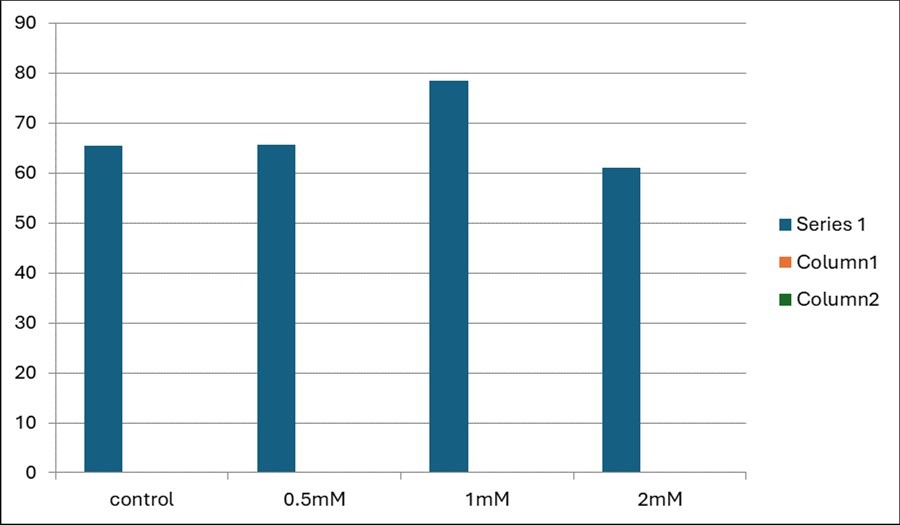
Download figure
Effect of glutamine and glycine addition during Vitrification on cleavage of vitrified/warmed cow oocytes:
The cleavage rate of cow oocytes after vitrification by using different treatments of glutamine and glycine was determined (Table 3, Figure 3). The highest cleavage rate was reported when using 1mM glutamine and glycine (21.57±4.09 a) which was significantly increased when compared to the control group (10.89±3.11 b). Addition of 0.5mM glutamine and glycine significantly increased cleavage rate when compared to the control group (12.12±3.30 b vs 10.89±3.11 b). Addition of 2mM glutamine and glycine significantly decreased cleavage rate when compared to the control group (08.00± 2.73 b vs 10.89±3.11 b).
Table 3. Effect of glutamine and glycine addition during Vitrification on cleavage of vitrified/warmed cow oocytes| Group | No. of oocytes | Cleavage mean ± SD (%) |
|---|---|---|
| Group 1 control | 101 | 10.89±3.11 b |
| Group2 0.5mM | 99 | 12.12±3.30 b |
| Group3 1mM | 102 | 21.57±4.09 a |
| Group4 2mM | 100 | 08.00± 2.73 b |
Figure 3. Effect of glutamine and glycine addition during vitrification on cleavage of vitrified/warmed cow oocytes. The data is presented as mean ± SD. Different letters a, b, c & d indicate a significant difference (P≤0.05) between treatment groups.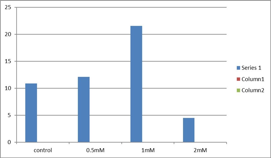
Download figure
Effect of glutamine and glycine addition during Vitrification on blastocysts of vitrified/warmed cow oocytes:
The blastocyst of cow oocytes after Vitrification by using different treatments of glutamine and glycine was determined (Table 4, Figure 4). The highest blastocyst rate was reported when using 1mM glutamine and glycine (09.80±2.96) which significantly increased blastocyst formation rate when compared to control (2.97±1.70). Addition of 0.5mM glutamine and glycine significantly increased this parameter when compared to the control group (3.03±1.73 b vs 2.97±1.70), but addition of 2mM result didn’t differ from control (3.00±1.71 b vs 2.97±1.70).
Table 4. Effect of glutamine and glycine addition during Vitrification on blastocysts of vitrified/warmed cow oocytes| Group | No. of oocytes | Blastocyst mean ± SD (%) |
|---|---|---|
| Group 1 control | 101 | 2.97±1.70 b |
| Group2 0.5mM | 99 | 3.03±1.73 |
| Group3 1mM | 102 | 09.80±2.96 a |
| Group4 2mM | 100 | 3.00±1.71 b |
Figure 4. Effect of glutamine and glycine addition during Vitrification on blastocysts of vitrified/warmed cow oocytes. The data is presented as mean ± SD. Different letters a, b, & c indicate a significant difference (P≤0.05) between treatment groups.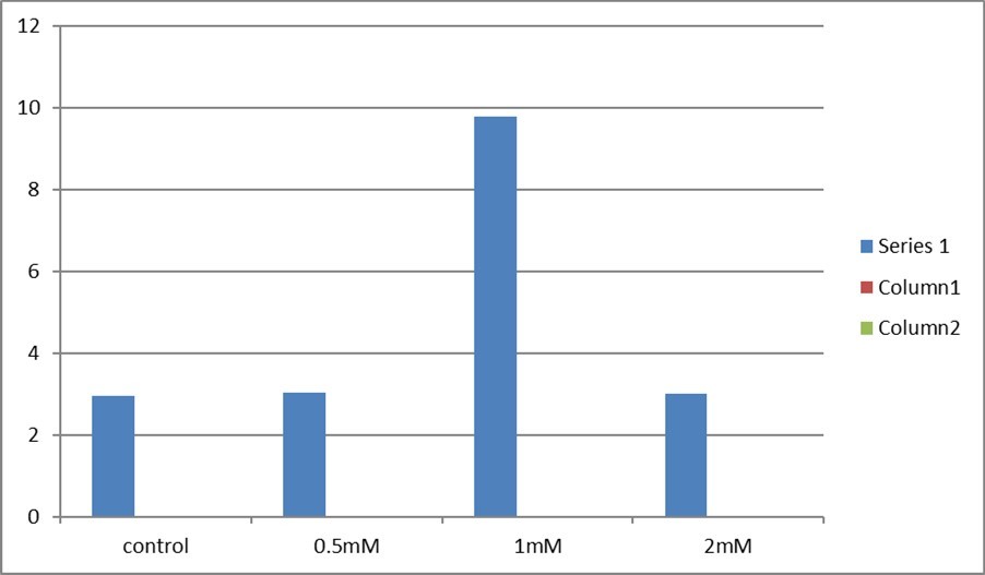
Download figure
Discussion
The effects of supplementation of cultural media with glutamine and glycine on the bovine oocytes’ morphology
Glycine, glutamate, and cysteine are the constitutive amino acids (CAA) involved in the production of GSH (Glu). GSH is produced in two ATP-dependent stages in mammalian cells. First, the enzyme γ-glutamylcysteine synthetase (GCL) converts Glu and Cys into γ-glutamylcysteine. Gly is then added to the dipeptide by glutathione synthetase (GS) 24. The limiting step in the synthesis of GSH is the reaction that GCL catalyzes. Because GCL is inactive, buthionine sulfoximine (BSO) is the most commonly used chemical to study GSH production25, 24. The three constitutive amino acids (CAA) glutamate (Glu), glycine (Gly), and cysteine (Cys) are combined to form glutathione (GSH), an antioxidant. In early embryo development, fertilization, and oocyte maturation, glutathione is essential. It was proven that GSH synthesis occurs during IVF. Furthermore, GSH levels reverted to control (mTALP) when BSO (mTALP + CAA + BSO) was added, inhibiting the increase in GSH seen in zygotes produced with mTALP + CAA medium. This is the first time that we are aware that GSH is produced during bovine insemination 26. In the present study, the supplementation of the IVM with 1mM glutamine and glycine resulted in a higher morphology rate than the control and other treatments.
Under in vitro culture conditions, gametes and embryos are subjected to elevated concentrations of reactive oxygen species (ROS) 27, 28, 29. ROS cause lipid peroxidation30, 31, DNA damage and even sperm cell death, sperm-oocyte fusion capacity, sperm motility reduction, and embryonic development delay 26. The primary non-protein sulfhydryl molecule in mammalian cells that has the capacity to shield cells from oxidative damage is glutathione (GSH). GSH affects disulfide reduction, DNA and protein synthesis, and amino acid transport, among other things 25, 32. Mammalian cells cannot synthesize cysteine from scratch, and the trans-sulfuration route required for cysteine production from methionine is only found in the liver and a few other tissues 33. Because mammalian cells can not maintain a considerable amount of cysteine, GSH may play an important role in regulating the cellular stress response during cysteine depletion34 .
The effects of supplementation of cultural media with glutamine and glycine on the bovine oocytes’ viability
Non-essential amino acids are essential for the body's regular physiological function and nutritional balance. Many non-essential amino acids, such as glutamine and glutamic acid, regulate fundamental metabolic operations of cells that are critical for animal survival, development, and reproduction 35.
The supplementation of the IVM with 1mM glutamine and glycine resulted in viability for the immature bovine oocytes than the other concentrations and control treatment. This may be attributed to the availability of CYS may be the rate-limiting factor for GSH synthesis during in vitro fertilization in cow oocytes during in vitro maturation 36. The γ-glutamyl cycle uses cysteine as a rate-limiting step in the synthesis of GSH 36, 37. It has been shown in HepG2 cells that, in the lack of amino acids, CYS deprivation alone might lower GSH levels, which could then be raised by CYS supplementation34.
Remarkably, throughout the last few years, new functions for glutathione (GSH) in proteins, apoptosis, signal transmission, gene expression, and nitric oxide (NO) metabolism have been identified 7, 33, 38. In order to get much-needed data regarding quantitative features of GSH synthesis and catabolism in the entire body and in particular cell types (such as erythrocytes), studies of in vivo GSH turnover in humans were most recently started 40, 41. The latest advancements in GSH metabolism and their effects on health and illness may illustrate the positive effects of a higher concentration of glutamate and glycine in the current study.
The effects of supplementation of cultural media with glutamine and glycine on the immature bovine cleavage rate
In the current study, we investigated the effects of glutamate and glycine supplementation to the IVM on the immature bovine cleavage rate. In the same line for the morphology and viability rates, the 1mM concentration of both amino acids is associated with a higher cleavage rate when compared to the other levels and control. This may be attributed to the vital role of both glutamate and glycine in the regulation of GSH synthesis 33
In brief, glutamate regulates GSH synthesis by two mechanisms: 1) cystine absorption and 2) avoidance of GSH inhibition of GCS. The system Xc amino acid transporter (8) is shared by glutamate and cysteine. Cysteine uptake is competitively inhibited by glutamate when extracellular glutamate concentrations are high, as in patients with advanced cancer, HIV infection, and spinal cord or brain injury, as well as in cell culture medium containing high levels of glutamate 42, 43. GSH is a nonallosteric feedback inhibitor of GCS, but it competes with glutamate for binding to the enzyme 44. GSH production is accelerated and its concentration is particularly high when intracellular glutamate concentrations are extremely high, as in canine erythrocytes 44.
On the other hand, Glycine availability may be decreased in response to protein deficiency, sepsis, and inflammatory stimuli 45, 46. When hepatic glycine oxidation is increased as a result of elevated glucagon levels or diabetes 47, this amino acid may constitute a limiting factor for GSH synthesis. Glycine availability restricts erythrocyte GSH synthesis in burned patients 41 and children recuperating from severe malnutrition 41. It is worth noting that dietary glycine supplementation raises hepatic GSH levels in protein-deficient rats challenged with TNF-45.
The effects of supplementation of cultural media with glutamine and glycine on the immature bovine blastocyst rate
Glutamine plays a crucial role in the biology of reproduction. Accordingly, we hypothesized that the supplementation of IVM media with glutamine may play a crucial role for improvement of the developmental competence of the immature bovine oocytes. The in vitro production of bovine embryos is crucial for medical and agricultural research. The medium modified synthetic oviduct fluid with amino acids (mSOFaa) is widely used for in vitro growth of bovine embryos 48. Glutamine is a necessary component of the mSOFaa fluid. Glutamine is a crucial source of energy for oocyte maturation and early embryonic evelopment 33, 49 and it is highly plentiful in follicular fluid 50, 51, compared to alanine and serine amino acid levels. Approximately 70% of glutamine is transformed to carbon dioxide during in vitro growth via the tricarboxylic acid cycle 52 . In addition to its role as an energy substrate, glutamine's use as an anaplerotic substrate to synthesize glutamate and then a-ketoglutarate is critical to a number of conversion steps in intermediary metabolism, including NAD conversion and the synthesis of heteropolysaccharides, glycoproteins, and other amino acids 49.
Glutathione is a tripeptide, -L-glutamyl-L-cysteinyl-glycine, that is found in all mammalian tissues but is most abundant in the liver. Glutathione is found in two forms: thiol-reduced (GSH) and disulfide-oxidized (GSSG)53. GSH is the most abundant type, present in most cells at millimolar quantities (liver 5-10mM). The content of GSSG is less than 1% of that of GSH54. GSH is stored in three primary reservoirs in eucaryotic cells. Almost 90% of cellular GSH is found in the cytosol, 10% in the mitochondria, and a trace in the endoplasmic reticulum 55, 56. The rat liver's cytosolic GSH turnover rate is rapid, with a half-life of 2-3 hours. GSH's peptide bond connects glutamate and cysteine via the -carboxyl group. The evidence suggests that dietary amino acid balance has a significant impact on protein nutrition and thus GSH homeostasis 33, 44, 57. The availability of sulfur-containing amino acids, as well as glutamate (glutamine or BCAAs) and glycine (or serine), is very important for optimizing GSH synthesis. Thus, GSH production is hindered in the erythrocytes of children with edematous protein-energy malnutrition and pigs with protein shortage, resulting in GSH deficit 32, 33. Increased urine excretion of 5-oxoproline, a -glutamyl cycle intermediate, is a helpful signal of decreased availability of cysteine and/or glycine for GSH synthesis in vivo 32, 33, 45.
In the current study, the supplementation of IVM media with 1mM of both amino acids showed a higher blastocyst rate of the immature bovine oocytes. It was proven that Glutathione (GSH) is a ubiquitous intracellular peptide that performs a variety of tasks such as detoxification, antioxidant defense, thiol status maintenance, and cell proliferation control. GSH is carefully controlled and produced in the cytoplasm of all mammalian cells 44 . The availability of cysteine, the sulfur amino acid precursor, and the activity of the rate-limiting enzyme glutamate cysteine ligase (GCL) are the key determinants of GSH formation. GCL is made up of a catalytic (GCLC) and a modifier (GCLM) subunit, both of which are regulated at different levels and at times differentially. GSH synthetase (GS), the second enzyme of GSH production, is also controlled in a coordinated manner as GCL subunits, and its up-regulation can further boost the cell's capacity to produce GSH 32, 57.
In conclusion the semiessential amino acid supplementation to the IVM media plays a crucial role in the improvement of the developmental competence of immature oocytes in bovine. Further studies are needed to investigate the molecular and biochemical effects of these amino acids on the immature oocytes either in vitro maturation and/or vitrification.
References
- 1.Y, M A Elmetwally, J G Maldonado-Estrada, F W Bazer. (2017) Physiological importance of polyamines. Zygote 25, 244-255.
- 2.Sánchez-Jiménez F, M Á, Villalobos-Rueda L, J L Urdiales. (2019) Polyamines in mammalian pathophysiology. Cell. Mol. Life Sci 76, 3987-4008.
- 3.M A Elmetwally, Li X, G A Johnson, R C Burghardt, C M Herring et al. (2022) Dietary supplementation with L-arginine between days 14 and 25 of gestation enhances NO and polyamine syntheses and the expression of angiogenic proteins in porcine placentae. Amino Acids 54, 193-204.
- 4.C M Herring, F W Bazer, G A Johnson, Seo H, Hu S et al. (2022) Dietary supplementation with 0.4% L-arginine between days 14 and 30 of gestation enhances NO and polyamine syntheses and water transport in porcine placentae. J. Anim. Sci. Biotechnol 13, 134-10.
- 5.Y, M A Elmetwally, Tang W, Satterfield C, Dunlap K et al. (2018) Functional roles of agmatinase during the peri-implantation period of pregnancy in sheep. Amino Acids 50, 293-308.
- 6.Moon H-S, Lee H-G, Chung C-S, Choi Y-J, Cho C-S. (2008) Physico-chemical modifications of conjugated linoleic acid for ruminal protection and oxidative stability. Nutr Metab (Lond) 5, 16-10.
- 7.Wu G. (2022) Nutrition and metabolism: foundations for animal growth, development, reproduction, and health. Adv. Exp. Med. Biol 1354, 1-24.
- 8.S M McKnight, R M Simmons, Wu G, M C Satterfield. (2020) Maternal arginine supplementation enhances thermogenesis in the newborn lamb. J. Anim. Sci 98, 10-1093.
- 9.Li P, Wu G. (2022) Important roles of amino acids in immune responses. The. British Journal of 127(3), 398-402.
- 11.K P Suresh, Nandi S, Mondal S. (2009) Factors affecting laboratory production of buffalo embryos: a meta-analysis. Theriogenology, 72(7). 978-985.
- 12.A-s A-H El-Naby, K G Mahmoud, Y F Ahmed, M E Abouel-Roos, A E. (2013) Effect of Season of the year and Ovarian Structures on Oocytes Recovery Rate, Quality and Meiotic Competence in Egyptian Buffaloes. Glob Vet 10, 408-412.
- 13.Mostagir A, Elmetwally M, Montaser A, Zaabel S. (2019) . Effects of L Carnitine and Cryoprotectants on Viability Rate of Immature Buffalo Oocytes in vitro After Vitrification. AJVS 62 45-10.
- 14.Abdeldayem M, Zaabel S, Badr M, Sharawy H, Mostagir A et al. (2022) Effects of L arginine supplementation on in-vitro maturation and cryotolerance of immature buffalo oocyte. Mans. Vet. Med. J 0, 0-0.
- 15.Abdulkarim A, Balboula A, Badr M, Bedir W, Zaabel S. (2021) Comparing in vitro maturation rates in buffalo and cattle oocytes and evaluating the effect of cAMP modulators on maturation and subsequent developmental competence. Mans. Vet. Med. J 22, 136-140.
- 16.G T Sharma, P K Dubey, Chandra V. (2010) Morphological changes, DNA damage and developmental competence of in vitro matured, vitrified-thawed buffalo (Bubalus bubalis) oocytes: A comparative study of two cryoprotectants and two cryodevices. Cryobiology 60, 315-321.
- 17.Martins R, Costa E, Chagas J, Ignácio F, Torres C et al. (2005) Effects of vitrification of immature bovine oocytes on in vitro maturation. Animal Reproduction 2, 128-134.
- 18.Nandi S, Ravindranatha B, Gupta P, Sarma. P.(2002b): Timing of sequential changes in cumulus cells and first polar body extrusion during in vitro maturation of buffalo oocytes. Theriogenology 57, 1151-1159.
- 19.AbdAllah S, Khalil A, Ali K. (2008) The use of Trypan blue staining to select the developmentally competent dromedary camel oocytes and its effect on in vitro maturation rate. The Proc. 20th Annual Scientific Conference 2-7.
- 20.A Z Balboula, Yamanaka K, Sakatani M, A O Hegab, S M Zaabel et al. (2010) Cathepsin B activity is related to the quality of bovine cumulus oocyte complexes and its inhibition can improve their developmental competence. Molecular Reproduction and Development: Incorporating Gamete Research. 439-448.
- 21.Parrish J, Susko-Parrish J, Leibfried-Rutledge M, Critser E, Eyestone W et al. (1986) vitro fertilization with frozen-thawed semen. Theriogenology 25, 591-600.
- 22.Hussien A, Sharawy H, Lenis Y, James D, Turna O et al. (2021) The impact of different estrus synchronization programs on postpartum holstein dairy cow reproductive performance. Mans. Vet. Med. J 22, 124-130.
- 23.M A Elmetwally, G E Elshopakey, Eldomany W, Eldesouky A, Samy A et al. (2021) Uterine, vaginal and placental blood flows increase with dynamic changes in serum metabolic parameters and oxidative stress across gestation in buffaloes. Reprod. Domest. Anim 56, 142-152.
- 24.O W Griffith, Meister A. (1979) Potent and specific inhibition of glutathione synthesis by buthionine sulfoximine (S-n-butyl homocysteine sulfoximine). The. Journal of Biological 254(16), 7558-7560.
- 25.M V Lafleur, J, Joenje H, E J Westmijze, Retèl J. (1994) The ambivalent role of glutathione in the protection of DNA against singlet oxygen. Free Radical Research, 21(1). 9-17.
- 26.J P Anchordoquy, R M Lizarraga, J M Anchordoquy, Nikoloff N, D E Rosa et al. (2019) Effect of cysteine, glutamate and glycine supplementation to in vitro fertilization medium during bovine early embryo development. Reproductive biology, 19(4). 349-355.
- 27.J C Deluao, Winstanley Y, R L, Pacella-Ince L, M B Gonzalez et al. (2022) . OXIDATIVE STRESS AND REPRODUCTIVE FUNCTION: Reactive oxygen species in the mammalian pre-implantation embryo. Reproduction, 164(6) 95-108.
- 28.Kalehoei E, Moradi M, Azadbakht M, Zhaleh H, Parvini M et al. (2022) In vitro maturation medium supplementation: utilization of repaglinide, L-carnitine, and mesenchymal stem cell-conditioned medium to improve developmental competence of oocytes derived from endometriosis mouse models. Brazilian. Journal of Medical and Biological Research, 55 11948.
- 29.M K Kim, J K Park, S K Paek, J W Kim, I P Kwak et al. (2018) Effects and pregnancy outcomes of L-carnitine supplementation in culture media for human embryo development from in vitro fertilization. The. Journal of Obstetrics and Gynaecology 44(11), 2059-2066.
- 30.Boni R, Gallo A, Cecchini S. (2017) Kinetic activity, membrane mitochondrial potential, lipid peroxidation, intracellular pH and calcium of frozen/thawed bovine spermatozoa treated with metabolic enhancers. Andrology, 5(1). 133-145.
- 31.Hamilton T R dos S, Castro L S de, Delgado J de C, Assis P M de, Siqueira A F P et al. (2016) Induced lipid peroxidation in ram sperm: semen profile, DNA fragmentation and antioxidant status. Reproduction, 151(4). 379-390.
- 32.Wu G, Fang Y-Z, Yang S, J R Lupton, N D Turner. (2004) Glutathione metabolism and its implications for health. The. Journal of 134(3), 489-492.
- 33.Wu Y, Zhang X, Bardag-Gorce F, Robel R C V, Aguilo J et al. (2004) Retinoid X receptor alpha regulates glutathione homeostasis and xenobiotic detoxification processes in mouse liver. Molecular. 65(3), 550-557.
- 34.Yu X, Y C Long. (2016) Crosstalk between cystine and glutathione is critical for the regulation of amino acid signaling pathways and ferroptosis. Scientific. Reports, 6 30033.
- 35.Wu G. (2014) Dietary requirements of synthesizable amino acids by animals: a paradigm shift in protein nutrition. Journal of animal science and biotechnology, 5(1). 34.
- 36.T C Sovernigo, P R Adona, P S Monzani, Guemra S, Barros F et al. (2017) Effects of supplementation of medium with different antioxidants during in vitro maturation of bovine oocytes on subsequent embryo production. Reproduction in domestic animals 52(4), 561-569.
- 37.Chance B, Sies H, Boveris A. (1979) . Hydroperoxide metabolism in mammalian organs. Physiological Reviews, 59(3) 527-605.
- 38.D M Townsend, K D Tew, Tapiero H. (2003) . The importance of glutathione in human disease. Biomedicine & Pharmacotherapy (Biomedecine & Pharmacotherapie),57(3–4) 145-155.
- 39.Badaloo A, Reid M, Forrester T, W C Heird, Jahoor F. (2002) Cysteine supplementation improves the erythrocyte glutathione synthesis rate in children with severe edematous malnutrition. The. American Journal of Clinical 76(3), 646-652.
- 40.Reid M, Badaloo A, Forrester T, J F Morlese, Frazer M et al. (2000) In vivo rates of erythrocyte glutathione synthesis in children with severe protein-energy malnutrition. American. Journal of Physiology. Endocrinology and 278(3), 405-12.
- 41.Yu Y-M, C M Ryan, Fei Z-W, Lu X-M, Castillo L et al. (2002) Plasma L-5-oxoproline kinetics and whole blood glutathione synthesis rates in severely burned adult humans. American. Journal of Physiology. Endocrinology and 282(2), 247-58.
- 42.Tapiero H, Mathé G, Couvreur P, K D Tew. (2002) . II. Glutamine and glutamate. Biomedicine & Pharmacotherapy (Biomedecine & Pharmacotherapie),56(9) 446-457.
- 43.Watford M. (2015) Glutamine and glutamate: Nonessential or essential amino acids?Animal Nutrition, 1(3). 119-122.
- 44.O W Griffith. (1999) Biologic and pharmacologic regulation of mammalian glutathione synthesis. Free. Radical Biology & Medicine, 27(9–10) 922-935.
- 45.R F Grimble, A, Persaud C, M J Wride, Delers F et al. (1992) Cysteine and glycine supplementation modulate the metabolic response to tumor necrosis factorαin rats fed a low protein diet. The. Journal of 122(11), 2066-2073.
- 46.Persaud C, Forrester T, A. (1996) Urinary excretion of 5-L-oxoproline (pyroglutamic acid) is increased during recovery from severe childhood malnutrition and responds to supplemental glycine. The. Journal of 126(11), 2823-2830.
- 47.G M Mabrouk, Jois M, J T Brosnan. (1998) Cell signalling and the hormonal stimulation of the hepatic glycine cleavage enzyme system by glucagon. The. Biochemical Journal, 330 ( Pt 2, 759-763.
- 48.Gutiérrez-Adán A, Lonergan P, Rizos D, F A Ward, M P Boland et al. (2001) Effect of the in vitro culture system on the kinetics of blastocyst development and sex ratio of bovine embryos. Theriogenology, 55(5). 1117-1126.
- 50.Y M Elhassan, Wu G, A C Leanez, R J Tasca, A J Watson et al. (2001) Amino acid concentrations in fluids from the bovine oviduct and uterus and in KSOM-based culture. 55(9), 1907-1918.
- 51.S A Hugentobler, M G Diskin, H J Leese, P G Humpherson, Watson T et al. (2007) Amino acids in oviduct and uterine fluid and blood plasma during the estrous cycle in the bovine. Molecular Reproduction and Development, 74(4). 445-454.
- 52.C L, R J Tasca, C A Ziomek. (1990) Glutamine uptake and utilization by preimplantation mouse embryos. in CZB medium. Journal of Reproduction and Fertility, 89(1) 335-346.
- 53.S C Lu. (1998) Regulation of hepatic glutathione synthesis. Seminars. in Liver Disease, 18(4) 331-343.
- 54.T P Akerboom, Bilzer M, Sies H. (1982) The relationship of biliary glutathione disulfide efflux and intracellular glutathione disulfide content in perfused rat liver. The. Journal of Biological 257(8), 4248-4252.
- 55.Dos Anjos Cordeiro, M J, L C Santos, B R Santos, E O Santos et al. (2023) Redox profile and mediators of the unfolded protein response (UPR) in the placenta of rats during pregnancy. Reproduction, fertility. and 35(10), 539-551.