
Retracted: Nutrient Foramen of Fibula in Relation to Distal end, Potential Implications for Vascularised Bone Graft Surgery
Abstract
Background
Free vascularised fibular bone grafting has gained popularity in various Orthopaedic and Oral & maxillofacial reconstructive surgeries. The objective of the present study was to identify the morphology and topography of nutrient foramina of fibula and to determine the foraminal index (FI) of the fibula using a more surgeon friendly bony landmark.
Methods
The study comprised examination of 100 fibulae specimens. Each bone was divided into 03 parts and topographical analysis was performed on each section. The nutrient foramina were identified macroscopically using size 24-gauge needle. Modified Hughes formula was used to calculate the foraminal index using distance of foramen (DF) from distal end which is easier to palpate in living human beings, total length of fibula (TL); and the formula was DF/TL x100.
Results
With respect to fibulae, 98% had single foramen and foramen was absent in 2%. The mean foraminal index (FI) was 56% for fibulae using modified Hughes’ formula. The majority of the fibulae showed nutrient foramen in the middle 3rd in relation to distal end of fibula.
Conclusion
The study provides information on the morphology of nutrient foramina in relation to easily palpable landmark on living human beings, which can provide guidance to surgeon while performing microvascular bone transfer procedures.
Author Contributions
Academic Editor: Abdelmonem Awad Mustafa Hegazy, Professor and Former Chairman of Anatomy and Embryology, Department, Faculty of Medicine, Zagazig University, Egypt.
Checked for plagiarism: Yes
Review by: Single-blind
Copyright © 2019 Amjad N Bhatti
 This is an open-access article distributed under the terms of the Creative Commons Attribution License, which permits unrestricted use, distribution, and reproduction in any medium, provided the original author and source are credited.
Competing interests
The authors have declared that no competing interests exist.
Citation:
Introduction
Vascularised bone grafting has become a treatment of choice in various reconstructive surgeries 1, 2, 3, 4. Knowledge of Anatomy of nutrient arterial blood supply to fibula is important for surgeon as it provides a useful adjunct in preoperative and intraoperative assessment to avoid potentially disastrous ischaemic complications after harvest of the flap. Clinical examination is often supplemented by other investigations. Postal survey results show most of surgeons relying on preoperative clinical examination with colour flow doppler in the initial assessment of lower limb’s vascularity followed by some kind of imaging studies 5.
The relatively invasive CT Angiography has the advantage of showing vascular anatomy but incidence of contrast allergy and nephrotoxicity are the associated risks. Magnetic resonance (MR) angiography has become increasingly popular and has the advantage of being non-invasive with comparable accuracy 5, however its availability in peripheral hospitals plus increased costs are limiting factors.
There is relative paucity of literature as respect to exact morphology and topography of diaphysial nutrient foramina in fibula. A literature search on Pubmed was carried out by Bolean method combining the terms ‘Diaphysial’ and ‘nutrient foramen’ and ‘Fibula’ which revealed only few such studies 6, 7, 8, 9, 10. In almost all these studies the nutrient foramen was calculated using proximal end of femur as reference point.
The aim of this review is to equip surgeon with back ground anatomical knowledge which is beneficial while operating for vascularised fibular bone graft considering a more easily palpable bony landmark during surgery.
Material and Methods
100 bone specimens were studied at our institute and King’s college London Institute; a size 24-guage needle was used to macroscopically identify nutrient foramen. In our study we calculated the foraminal index (FI) taking lower end of fibula or lateral malleolus as reference point (Figure 1). The reason for using distal end of fibula instead of proximal end is due to the fact of increasing level of obesity in these patients and potential difficulty encountered by surgeon due to various body habitus factors 11. The distal end is easier to palpate in living human beings as it is more prominent and mainly subcutaneous.
Figure 1.A fibula specimen and materials used in methods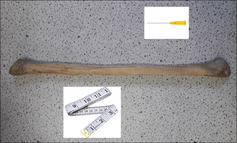
Results
Out of 100 bone specimens studied, 98 showed definite nutrient foramen confirmed by needle insertion, 02 bones did not show the nutrient foramen. The reason could be ossification, due to lack of availability of forensic data for these specimens we are not sure about the exact age of these bone specimens. The mean total length of fibular specimens was found to be 35.41 cm, mean foraminal distance from distal end of fibula was 19.69 cm with mean Foraminal index (FI) using modified Hughe’s formula FI =DF/TL× 100 (DF stands for distance of nutrient foramen from distal end and TL stands for total length) was 56% with standard deviation of 0.071.
Discussion
The fibula is supplied from one or more branches of the peroneal artery. In the present study, we observed the single fibular foramen in 98% of the cases, which is similar to the reports of Zahid et al 6, Campos et al 12 and McKee et al 13.
In study by Zahid et al 6, 85 bones (96.6%) of right side and 79 bones (98.8%) of left side had a single nutrient foramen. Mean foramen index was 47.6 for left and 50.2 on the right side. In the Korean study by Choi et al 14, the location of the nutrient foramen was found to be just proximal to the midpoint. The study by Kocabiyik et al 8 also found most nutrient arteries were near the middle third of the fibula. In study by Gumusburun et al 10, the common location for nutrient artery foramen is middle third of fibula 98.0 %. The mean foraminal index of the fibula was found to be 48.14 %. Campos et al 12 studied the nutrient foramina of both upper and lower limb long bones in Spanish specimens and found that in the fibula the nutrient foramen index is between 35 and 67 %. According to the Turkish study from Kizilkanat et al 9, foramina were located on the diaphysis and calculated Foraminal index between 26–83% for the fibula. In the present study, it was observed that the foramina were located on the diaphysis. The majority (90%) of the fibular specimens, foramina were located at the middle 3rd of fibula. These findings are almost similar to the previous reports from other studies; however, these mentioned studies calculated the foraminal index using proximal end of fibula as reference point.
The absence of nutrient foramina in long bones is well known 7, 8. It was reported that in instances where the nutrient foramen is absent, the bone is likely to be supplied by periosteal arteries. In the present study, the nutrient foramen was absent in 02 bone specimens however due to lack of availability of forensic data we are not sure if it represents age related ossification.
Our study has some potential limitations. These include age and sex differences which were not considered as we were not able to estimate the age and gender of the bones studied. The present study could not address these factors as the study involved cadaver dry bones without availability of forensic data. These differences might alter the results as the anatomy of foramina might differ according to gender and age.
Conclusion
The present study provides valuable information on the fibular bone nutrient artery foramina using an easily palpable bony landmark. As techniques such as microvascular bone transfer are becoming more popular, information relating to the anatomical description of these foramina is vital to preserve the circulation of affected bony structures. Use of a bony land mark which is easily palpable during surgery could be helpful for surgeon.
Acknowledgment
Authors would like to acknowledge the contributions of Lydia Carline Anatomy Prosector at Brighton & Sussex Medical School and Kirsty Massetti at Kings College London for their valuable support in providing access to Fibular bone specimens.
References
- 1.Yamamoto N, Morikawa T, Yakushiji T, Shibahara T. (2018) Mandibular Reconstruction with Free Vascularized Fibular Graft. The Bulletin of Tokyo Dental College. 59(4), 299-311.
- 2.Liu S, Tao S, Tan J, Hu X, Liu H et al. (2018) Long-term follow-up of fibular graft for the reconstruction of bone defects. , Medicine (Baltimore) 97(40), 12605.
- 3.Makiguchi T, Yokoo S, Hashikawa K, Miyazaki H, Terashi H. (2015) Evaluation of bone height of the free fibula flap in mandible reconstruction. , J Craniofac Surg 26(3), 673-6.
- 4.Matsuura M, Ohno K, Michi K, Egawa K, Takiguchi R. (1999) Clinicoanatomic examination of the fibula: anatomic basis for dental implant placement. , Int J Oral Maxillofac Implants 14(6), 879-84.
- 5.Whitley S P, Sandhu S, Cardozo A. (2004) Preoperative vascular assessment of the lower limb for harvest of a fibular flap: the views of vascular surgeons in the United Kingdom. , British Journal of Oral and Maxillofacial Surgery 42(4), 307-10.
- 6.Zahid A, Shakir M A, Shireen R. (2015) Morphological and Topographical Anatomy of Diaphyseal Nutrient Foramina of Dried Pakistani Fibulae. , Journal of the College of Physicians and Surgeons--Pakistan: JCPSP 25(8), 560-3.
- 7.Murlimanju B, Prashanth K, Prabhu L V, Chettiar G K, Pai M M et al. (2011) Morphological and topographical anatomy of nutrient foramina in the lower limb long bones and its clinical importance. The Australasian medical journal. 4(10), 530-7.
- 8.Kocabiyik N, Yalcin B, Ozan H. (2007) Variations of the nutrient artery of the fibula. , Clin Anat 20(4), 440-3.
- 9.Kizilkanat E, Boyan N, Ozsahin E T, Soames R, Oguz O. (2007) Location, number and clinical significance of nutrient foramina in human long bones. , Annals of anatomy = Anatomischer Anzeiger : official organ of the Anatomische Gesellschaft 189(1), 87-95.
- 10.Gumusburun E, Adiguzel E, Erdil H, Ozkan Y, Gulec E. (1996) A study of the nutrient foramina in the shaft of the fibula. Okajimas folia anatomica Japonica. 73(23), 125-7.
- 11.Wang Y C, McPherson K, Marsh T, Gortmaker S L, Brown M. (2011) Health and economic burden of the projected obesity trends in the USA and the UK. The Lancet. 378(9793), 815-25.
- 12.Campos F F, Pellico L G, Alias M G. (1987) Fernandez-Valencia R. A study of the nutrient foramina in human long bones. Surgical and Radiologic Anatomy. 9(3), 251-5.
Cited by (2)
This article has been cited by 2 scholarly works according to:
Citing Articles:
Indian Journal of Orthopaedics (2023) Crossref
S. Das, Chandraprabha Choudhary, M. Mishra - Indian Journal of Orthopaedics (2023) Semantic Scholar
Indian Journal of Orthopaedics (2023) OpenAlex