Abstract
Molecular imaging is a new method in examining physiological studies in molecular dimensions. Among the various methods that have been introduced for this purpose, the magnetic resonance spectroscopy (MRS) method has made it possible to more accurately study the activities of the brain region as well as tumors in different parts of the body. MRS imaging is a type of non– invasive imaging technique that is used to study metabolic changes in the brain, stroke, seizure disorders, Alzheimer's disease, depression and also metabolic changes in other parts of the body such as muscles. In fact, since metabolic changes in the human body appear faster than anatomical and physiological changes, the use of this method can play an important role in the early detection and diagnosis of cancers, infections, metabolic changes and many other diseases.
Graphical abstract: CERN Large Hadron Collider (LHC) radiation source for magnetic resonance biospectroscopy in metabolic and molecular imaging and diagnosis of cancer.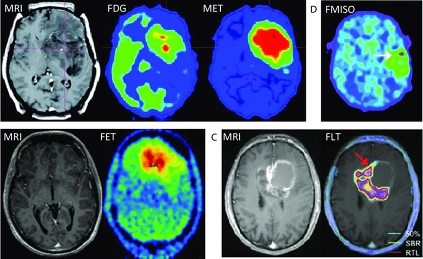
Author Contributions
Academic Editor: Xiaoyong Lu, Department of Chemistry and Biochemistry, Ohio University, Athens, Ohio, 45701USA
Checked for plagiarism: Yes
Review by: Single-blind
Copyright © 2022 Alireza Heidari, et al.
 This is an open-access article distributed under the terms of the Creative Commons Attribution License, which permits unrestricted use, distribution, and reproduction in any medium, provided the original author and source are credited.
Competing interests
The authors have declared that no competing interests exist.
Citation:
Introduction
MRI (MR) imaging is primarily related to the production of anatomical images, while in the MRS method, instead of an image, we will have a spectrum of the range of MR signals according to their intensity frequency (in Hertz or ppm) 1, 2, 3, 4, 5, 6, 7, 8, 9, 10, 11, 12, 13, 14, 15, 16, 17, 18, 19, 20, 21, 22, 23, 24, 25, 26, 27, 28, 29, 30, 31, 32, 33, 34, 35, 36, 37, 38. The signals recorded by MRI are mainly from protons in water and fat. In MRS studies other than hydrogen nucleus, other nuclei such as 31P, 7Li, 19F, 23Na and 13C have been used, which contain physiological information. By comparison, MRS aims to analyze the chemical composition of tissues in a very small number of much larger voxels 39, 40, 41, 42, 43, 44, 45, 46, 47, 48, 49, 50, 51, 52, 53, 54, 55, 56, 57, 58, 59, 60, 61, 62, 63, 64, 65, 66, 67, 68, 69, 70, 71, 72, 73, 74, 75, 76. The signal–to–noise ratio in MRS is lower than in MRI, therefore, the volume of selected voxels is considered larger for MRS. MRI removes chemical shift information, while the purpose of MRS is to enhance this information qualitatively and quantitatively 77, 78, 79, 80, 81, 82, 83, 84, 85, 86, 87, 88, 89, 90, 91, 92, 93, 94, 95, 96, 97, 98, 99, 100, 101, 102, 103, 104, 105, 106, 107, 108, 109, 110, 111, 112, 113, 114.
For cancer treatment, it is critical to be able to identify key biomolecules and molecular changes associated with cancer and harmful things, as well as to monitor the medically beneficial results against these targets. People who work to find information and doctors now have new tools to improve most aspects of cancer care thanks to recent developments in molecular imaging based on magnet–based (MR) methods. The broad definition of molecular imaging is "imaging techniques for detecting molecular signatures at the cellular and expression (tiny chemical assembly instruction inside of living things) levels. “This article discusses the (possible power or ability within/possibility of) these ways of doing things in improving medicine–based cancer care and reviews both established and newly appearing molecular MR methods in cancer–related medical care. It also talks about how molecular MR, as well as other ways of doing things with functional MR imaging (related to what holds something together and makes it strong), paves the way for custom–designed cancer treatment (success plans/ways to reach goals) 115, 116, 117, 118, 119, 120, 121, 122, 123, 124, 125, 126, 127, 128, 129, 130, 131, 132, 133, 134, 135, 136, 137, 138, 139, 140, 141, 142, 143, 144, 145, 146, 147, 148, 149, 150, 151, 152
Breast cancer is a common disease that affects women. It is the second leading factor in women's cancer–related deaths. Related to food processing and use), reprogramming takes place during the growth of cancer, sudden, unwanted entry into a location, and disease spread throughout the body. Body–structure–related and molecular processes have shown (possibility of/possibility of happening of) illustrating body–structure–related and molecular processes changes before (related to body structure) visible signs on ordinary MR imaging, as shown by functional magnet–based (MR) methods containing/making up an organized row of ways to do things. One of these is in vivo proton (1H) MR spectroscopy (MRS), which is widely used to distinguish breast cancer from other diseases by measuring compounds that contain more choline. In addition, the understanding of glucose and phospholipid (chemically processing and using food) was enhanced by the utilization of hyperpolarized 13C and 31P MRS. In vitro bright and sharp NMR spectroscopy and bright and sharp magic angle spectroscopy (HRMAS) can also be used to closely examine medical samples and examples (unharmed and in one piece tissues, tissue extracts, and various biofluids such as blood, urine, nipple breathes/inhales, and fine needle breathes/inhales) to gather information about the (related to processing and using food) body functions of living things. In addition to providing a deeper understanding of cancer (study of living things/qualities of living things) and chemically processing and using food, such studies can provide information on more metabolites than seen by in vivo MRS. The tumor subtypes were classified after a large number of NMR data sets related to ghosts or rainbow colors were analyzed using multivariate methods related to studying numbers. It demonstrated significant (possible greatness or power) progress in the creation of novel medically beneficial strategies. By putting into numbers (related to what holds something together and makes it strong), vasculature, diffusion, perfusion, and (related to processing and using food) (things that are different from what is usually expected) in vivo, multiparametric MRI approaches were found to be helpful in explaining how a disease works, particularly cancer. This review focuses on how NMR, MRS, and MRI can be used to understand breast cancer (study of living things and their qualities), identify a disease or problem or its cause, and monitor breast cancer in a way that is helpful to medicine 1.
Results and Discussion
MR spectroscopy analyzes molecules such as hydrogen ions or protons. Proton spectroscopy is more common. There are several metabolites or metabolic products that can be measured to differentiate between tumor types: Lactate or Lac N–acetyl aspartate or NAA Choline or Cho Creatine or Cr Myo–inositol or Myo Glutamate and Glutamine or Glx Lipid. The abundance of these metabolites is measured in units called parts per million (ppm) and plotted as peaks of different heights on the graph. The horizontal axis of the spectrum indicates the amount of chemical shift of each of these materials and the vertical axis indicates the amount of this chemical shift, which is the same signal resulting from the magnetic intensification of the core. By measuring the PPM of each of the mentioned metabolites and comparing them with normal brain tissue, neurologists can determine the type of tissue present. MR spectroscopy can be used to determine the type of tumor and whether it is malignant or benign, etc. Simultaneously with the discovery of MRI, the chemical shift effect was also identified. Chemical shift (chemical shift) is the basis of MRS. The origin of this effect is the response of the electrons of a molecule to the magnetic field 115, 116, 117, 118, 119, 120, 121, 122, 123, 124, 125, 126, 127, 128, 129, 130, 131, 132, 133, 134, 135, 136, 137, 138, 139, 140, 141, 142, 143, 144, 145, 146, 147, 148, 149, 150, 151, 152. In the MRI discussion, the nucleus or proton is affected by an external field with intensity B0 and therefore rotates around the field with the Larmor frequency, but the electrons themselves also create a protective effect or shield around the proton or nucleus, which is called the shielding constant. we say. The greater the electron cloud and the number and characteristics of the electronegativity, the greater this protection is, and therefore the nucleus does not see the actual external value of the field, so we expect hydrogens that are in tissues with less electron shielding to see a greater external magnetic field and according to the Larmor relation They rotate faster around the external field, while for tissues such as fat, where hydrogen protons have stronger bonds with carbons and electron shields, they rotate slower with the Larmor frequency 153, 154, 155, 156, 157, 158, 159, 160, 161, 162, 163, 164, 165, 166, 167, 168, 169, 170, 171, 172, 173, 174, 175, 176, 177, 178, 179, 180, 181, 182, 183. In fact, different metabolites have different hydrogen bonds and considering that the chemical shift in them differs according to what was mentioned, we can use it in Spectroscopy. In general, two different approaches are used in proton spectroscopy: Single voxel method that uses a sequence of STEAM or PRESS pulses and spectroscopic imaging methods that are also known as chemical shift imaging or CSI. In the first attempts to perform spectroscopic imaging, which is also referred to as MRS, the one– dimensional method was performed using phase coding in one direction. By using MRSI coding gradients, the phase methods in two directions were extended to two dimensions and subsequently to three dimensions with three–dimensional coding, which are called chemical shift imaging (Figure 1).
Figure 1. The phase methods in two directions were extended to two dimensions and subsequently to three dimensions with three–dimensional coding using MRSI coding gradients.
While most single voxel studies are performed in short TEs. MRSI studies are performed in long TEs. Low TE spectra contain the signal of a greater number of compounds and as a result better SNR, but their contamination with water and fat is also more. In contrast, high TE spectra have lower SNR, less visible compounds and different T2–weight values, but they have spectra with more separated resonances and a smoother background. The choice of method depends on the information needed in a specific medical or research application. For example, if spectroscopy is used to find the location of a stroke or seizure center in the brain, the microscopic extent of tumors and the intensity of tumor invasion in the prostate and brain, the CSI method is preferable because it is able to create a map of the amount of metabolites in order to diagnose lesions. Scattered to be used in different places. But if the tissue is studied in order to check the composition change at a specific point, the single voxel spectroscopy method will be the chosen method (Figure 2).
Figure 2. Schematic of single voxel spectroscopy method.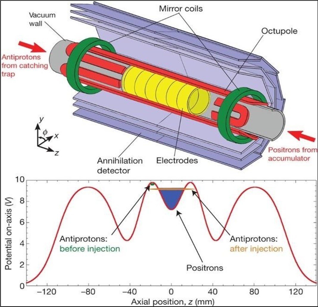
It is a non–invasive method. It can be used to monitor the chemical changes of tissues. We can simultaneously evaluate several metabolites. Two examples of where MRS is very helpful in the brain: The invasion of the tumor (Glioblastoma multiform (GBM) into the surrounding tissues, which is not clear in normal T2 images, but can be determined by MRS. By MRS, it is possible to distinguish two types of lesions that look similar to each other in normal MRI images (such as tumor recurrence and tumor necrosis after radiotherapy). MRS imaging has found wide applications in the field of cancer diagnosis. Among the fields of clinical application of MRS, we can mention the diagnosis (between normal and cancerous tissue, different types of cancer and neoplastic from non–neoplastic), designing the best treatment regimens for each patient, and monitoring the patient after treatment. MRS in tumors: In brain tumors, spectroscopy can determine the degree of malignancy. As malignancy increases, NAA and creatine decrease and choline, lactate and fat increase. Fat is seen in the necrotic parts of the tumor. Lactate concentration increases in rapidly growing tumors due to anaerobic glycolysis. Diagnosing tumor recurrence from the effects of radiotherapy: Increased choline is a marker for tumor recurrence. Changes due to radiotherapy usually decrease NAA, creatine and choline. If necrosis has occurred as a result of radiotherapy, fat and lactate can also be seen in the spectrum. Molecular imaging using spectroscopy Cerebral ischemia and infarction: When the brain suffers from ischemia, anaerobic respiration of glucose is used and lactate increases. Choline increases and NAA and creatine decrease. If it happens after ischemia, the fat signal is also seen. trauma: It is a useful method to assess the degree of nerve damage and predict the results. The clinical consequences are opposite to the NAA/Cr ratio, and the observation of lactate and fat indicates the seriousness of the condition. infectious diseases: decrease naa Inside the abscess, lactate, alanine, cytosolic acid and acetate increase. Alzheimer: In the advanced stages of Alzheimer's, NAA decreases and myo–inositol increases. MS: The increase of choline and lactate has shown that the increase of choline can be due to the increase of phospholipid as a result of breaking the myelin of the cell and the increase of lactate is due to the increase of the anaerobic respiration of the cell due to the increase of the cell metabolism. In addition, there is evidence of increased lipids, and most importantly, decreased NAA, which is caused by nerve damage. And recently, it has been found that glutamate and myoinositol levels increase in acute MS lesions. Parkinson: In most studies in Parkinson's disease, no changes in metabolites have been observed, only when Parkinson's has caused brain atrophy, a decrease in NAA in the basal ganglia has been observed (Figure 3, Figure 4, Figure 5, Figure 6).
Figure 3. Different spectra metabolites in different areas of the human body.
Figure 4. Infiltrating macrophages of cancer cells in interaction with hypoxia acidic pHe substrate deprivation.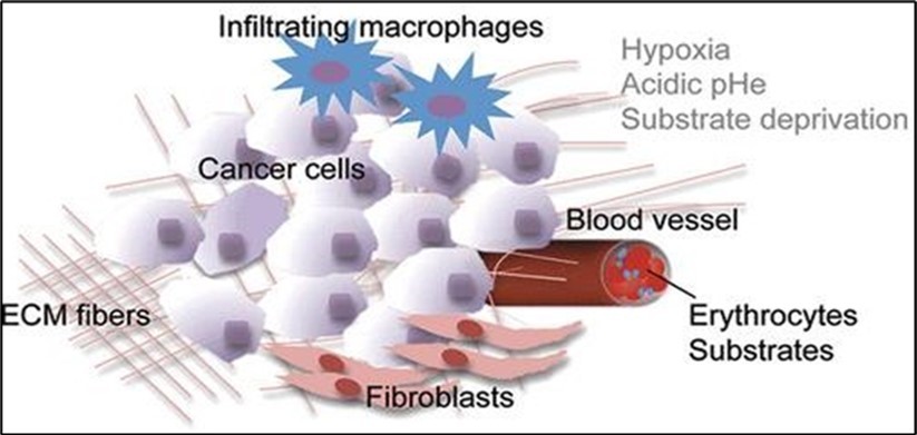
Figure 5. Schematic of different steps of CERN Large Hadron Collider (LHC) radiation source for magnetic resonance biospectroscopy in metabolic and molecular imaging and diagnosis of cancer.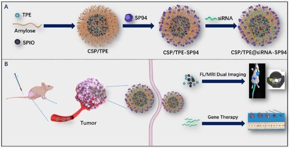
Figure 6. Simulation of CERN Large Hadron Collider (LHC) radiation source for magnetic resonance biospectroscopy in metabolic (left) and molecular (right) imaging and diagnosis of cancer.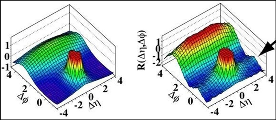
Conclusion, Summary, Outlook and Future Directions
MRS imaging method is a new method in molecular imaging that can be used in different types of differential diagnoses. Among the areas of clinical application of MRS, we can mention the diagnosis (between normal and cancerous tissue, different types of cancer and neoplastic from non–neoplastic), designing the best treatment regimens for each patient, and monitoring the patient after treatment. This method can solve the lack of ability of MRI method in examining pathology.
Measurements of molecular and cellular processes, such as the chemical processing and use of food, cell death, cell growth and spread, and biosynthetic pathways of various metabolites in vivo in cancer, can be made using molecular MR imaging. Every aspect of cancer–related medical care, including early disease detection, identification of a disease or problem or its cause, staging, personalized treatment, and treatment monitoring/supervision, can benefit from molecular imaging. Ovarian, lung, and male reproductive gland cancer are just a few of the many types of cancer for which molecular imaging had a significant impact on patient care. Detecting and curing disease in its most treatable phase, as well as saving a large number of lives, may be possible with molecular imaging's ability to detect (things that are different from what is usually expected) very early in the (development or increase over time/series of events or things) of disease. This could shift medicine away from causing reactions from other people or chemicals and toward preventing problems before they occur. In clinical arrangement, sub–atomic X–ray will make ready toward a major improvement in early discovery of illness, treatment arranging and watching/overseeing the restoratively supportive outcomes.
This survey momentarily introduced the (conceivable power or capacity inside/probability of) X–ray and MRS–based techniques in figuring out bosom malignant growth (investigation of living things/characteristics of living things) and the job of various MR biomarkers in illness (recognizable proof of a sickness or issue, or its goal), (proclamation about a potential future occasion), (looking at and testing so a choice can be made), medicinally supportive watching/managing, and cancer (rehashing occasion).Numerous metabolites were found in breast cancer patients through in vitro bright and sharp NMR studies of tissue extracts, nodes, serum, and blood plasma samples. More than two, but not many, metabolites, membrane metabolites like tCho and GPC, and amino acids like Ala, Glu, Gln, Lys, His, Gly, Ser, and Tau, as well as legal and law–based machines, methods, and ways, were shown to have changed in response to the changes. In addition, these metabolites could be used as disease–specific and prediction– related biomarkers in the treatment of breast cancer.
The molecular mixed–up nature of tumors was also connected to the mixed–up nature of tumors, which was related to food processing and use. However, a comprehensive and thorough description of the mixed–up nature of breast cancer (damage to body parts) is required in relation to food processing and use. X–ray and MRS are currently being utilized as (partner/helping) approaches to getting things done to clinical bosom tests, histology, and alternate approaches to getting things done. Information on tumor cellularity, perfusion, and stiffness are provided by MRI, which combines them to produce something superior. RI has emerged as an important tool for (determining the value, quantity, or quality of) the population of women at high risk over the past few years. The use of MRI in the detection of cancers that are occult on a mammogram has been demonstrated in numerous studies. However, due to its technical difficulties, breast MRS is still not performed regularly. MRS’s sensitivity is also constrained by a number of technical factors. However, recent computer and scientific advancements, like improving the design and sensitivity of breast coils and high–field MR systems, may be able to enhance the breast MRS's quality of being very close to the truth or true number. Even though the methods of MRI and MRS showed or told about a lot of biomarkers as potential candidates, they are only used in research labs at this time for (more than two, but not a lot of) reasons like technical difficulties and higher costs for procedures, equipment not being available, etc. For these markers to be used in clinics to provide decorated (with a personal touch) health care, they need to be developed with greater reproducibility.
Using MR techniques, it is necessary to demonstrate various histological types of breast cancer for a comprehensive understanding of its mixed nature. The ability of these methods to identify diseases may improve as a result of this. In addition, there is a requirement for simple, automated acquisition, learning, and post–processing sets of computer instructions that can be visualized (in your mind) and converted into numbers for Cho in tumors of a small size. The cost of MR procedures for more applications should be the primary focus of future research. Additionally, multi–center studies on the application of MRI and MRS strategies in medicine–based settings are required to "combine" them into a single unit. NMR spectroscopy of biofluids in women at risk for (related to things you get from your parents' genes) is also necessary to (figure out the worth, amount, or quality of). This is a potential area for future research that could aid in the classification of women at high risk for cancer and provide an early indication of the vulnerable population. In addition, it is crucial to carry out metabolomics studies in a well–organized manner in order to discover robust and healthy biomarkers for the (identification of a disease or problem, or its cause), as well as the outlook for the disease. The results of metabolomics research ought to be translated into the development of overly straightforward methods that could be easily implemented in medicine–based settings with low–cost effects, recommendations, and results. Long/big multi–center (acts of asking questions and attempting to find the truth about something) is required by recent methods like MR elastography. Utilizations of radiomics need to be thoroughly investigated, and X–ray practitioners need to gain a better understanding of the fundamental concepts, the creation of reproducible (done or made to look the same every time) sets of computer instructions, and the sharing of data for medicine–based applications.
Acknowledgements
This study was supported by the Cancer Research Institute (CRI) Project of Scientific Instrument and Equipment Development, the National Natural Science Foundation of the United Sates, the International Joint BioSpectroscopy Core Research Laboratory (BCRL) Program supported by the California South University (CSU), and the Key project supported by the American International Standards Institute (AISI), Irvine, California, USA.