
An Anatomical Study of the Tibia in the North Indian Population
Abstract
Introduction
The human tibia is a complex anatomical unit and the knowledge of its morphometric values is important in Forensic, Anatomic and Radiological cases in order to identify unknown bodies and stature.
Objective
It was to analyze the tibia, its morphometry, side difference and to investigate the position of nutrient foramina in tibia.
Methods
In this study, 60 adult human tibias (30 right and 30 left) were obtained from the Department of Anatomy SMSR SHARDA UNIVERSITY. In the study a total of two parameters i.e. Cross Section Index in the middle and Cnemicus Index of the bones were obtained and evaluated by using two instruments- a Measuring Tape and a Vernier Calipers. All the bones were dry and showed normal anatomical features.
Results
The mean Cross Section Index in the middle was calculated as 80.42 ± 11.33 on the right side and 78.15± 12.78 on the left side; and the Cnemicus index was 78.40 ± 13.19 on the right side and 70.84 ± 11.38 on the left side
Conclusion
The two parameters in the North Indian population were compared with other populations. The values were found to be almost comparable however there were subtle differences between different populations. The position of nutrient foramen was also assessed. This knowledge will thus help further researchers and orthopedic surgeons in various procedures like joint replacement therapy, fracture repair, bone grafts and vascularized bone microsurgery as well as in medico‐legal cases.
Article Information
- Received
- Accepted
- Published
Academic Editor: Abdelmonem Awad Mustafa Hegazy, Professor and Former Chairman of Anatomy and Embryology Department, Faculty of Medicine, Zagazig University, Egypt.
Checked for plagiarism: Yes
Review by: Single-blind
Copyright © 2019 Tiwari A, et al.
 This is an open-access article distributed under the terms of the Creative Commons Attribution License, which permits unrestricted use, distribution, and reproduction in any medium, provided the original author and source are credited.
Corresponding author: Aman Tiwari, , MBBS II Year, School of Medical Science & Research, Sharda University, Greater Noida —
Competing Interests
The authors have declared that no competing interests exist.
Funding
No specific funding statement was provided by the authors.
Data Availability
No data-availability statement was provided by the authors.
Citation:
Introduction
The tibia is situated at the medial side of the leg; and excepting the femur, is the longest bone of the skeleton 1. The anterior border of the tibia is the most prominent border. The anterior border and the adjacent medial surfaces are subcutaneous throughout their length and are commonly known as “shin” 2.
The tibia ossifies from three centers, one in the diaphysis and one at each proximal and distal epiphysis. The medial malleolus is merely an extension from the distal epiphysis 3.
The nutrient artery is the main source of blood supply to a long bone which enters the bone through the nutrient foramen. This artery is very important for the growth of bone in embryo and fetus as well as during the early ossification phase of bone 4.
Study of comparative association between length of bone and distance of nutrient foramen from either end is beneficial in calculating the length of a long bone which is significant in medico-legal and anthropometry work. From the length of the bone, the stature of an individual can also be found out 5.
Also, an understanding of the location and the number of the nutrient foramina in long bones is vital in various orthopedic surgical procedures such as joint replacement therapy, fracture repair bone grafts and vascularized bone microsurgery 6.
Hence, the present study was carried out to measure various dimensions of tibia to calculate the Cross-Section and Cnemicus Indices in an endeavor to provide a base line data pertaining to morphometric details of tibia with reference to knee arthroplasty in the North Indian population. The results of the study assume a special importance in view of technical advances in reconstructive surgical procedures in orthopedic practice.
Material and Methods
The present study was carried out on 60 tibias, 30 each from right and left side. Damaged and broken bones were not included. The following parameters of each bone were measured.
Cross-Section Index in Middle = (Transverse diameter in middle of bone/Maximum diameter in middle of bone) X 100
In this formula, transverse diameter in middle of the bone is calculated as the straight distance from the medial tibial border to the interosseous crest at the middle of the bone.
Maximum diameter in middle of the bone measures the straight distance of the anterior crest from the posterior surface in the middle of the bone.
Cnemicus Index= (Transverse diameter at level of Nutrient foramen/ Sagittal diameter at level of Nutrient Foramen) X100
In this formula, transverse diameter of the bone is calculated as straight distance from the medial tibial border to the interosseous crest at the level of the nutrient foramen.
Sagittal diameter in the middle of the bone measures the straight distance of the anterior crest from the posterior surface at the level of the nutrient foramen.
Location and number of Nutrient Foramina.
Two instruments were used in the study; a Vernier Caliper and Measuring Tape. Using these two instruments, three readings were obtained for each parameter on each bone and the mean of those three readings was taken.
The photographs were taken using a digital camera. The means and standard deviations of the entire data were calculated. The comparison of various dimensions of the right and left sides was performed and statistically analyzed.
Results
The mean Cross Section Index in the middle was calculated as 80.42 ± 11.33 on the right side and 78.15± 12.78 on the left side and the Cnemicus index was 78.40 ± 13.19 on the right side and 70.84 ± 11.38 on the left side (Table 1). A comparison in the mean values of the bone on the two parameters was made with other studies (Table 2 and Table 3). In one of the tibias there was presence of two nutrient foramens instead of the usual one. This was an unusual finding we observed during the study (Figure 1 and Figure 2). The most common location of the foramen was seen on the posterior surface in the upper one third of the bone.
Table 1. Showing Means and Standard Deviations (SD) of the two parameters of Tibia| PARAMETER | MEAN ± SD | |
| CROSS SECTION INDEX IN THE MIDDLE (cm) | RIGHTLEFT | 80.42 ± 11.3378.15 ± 12.78 |
| CNEMICUS INDEX (cm) | RIGHTLEFT | 78.40 ± 13.1970.84 ± 11.38 |
Figure 1. showing two nutrient foramina on shaft of tibia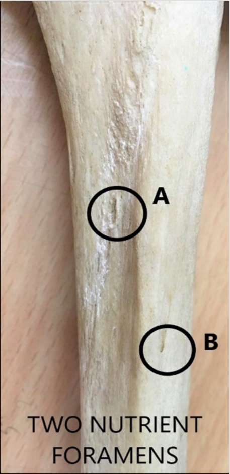
Download figure
Figure 2. Measurements done on tibia using Vernier caliper and measuring tape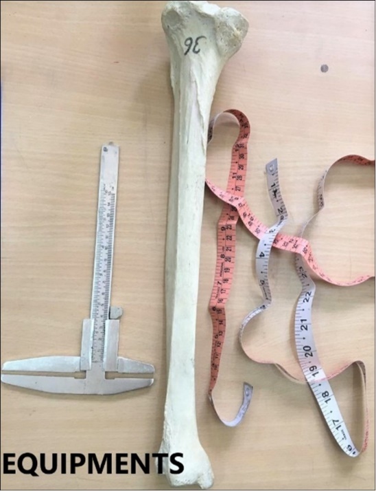
Download figure
Discussion
The vertically oriented tibia lies medial to, and is stronger than, the accompanying fibula. The tibia's proximal end, the tibial plateau, provides a surface for articulation with the femur, thereby allowing transmission of the body's weight as well as ground reaction forces. When both forces are transmitted strongly, as in jumping from an elevated position, the knee joint and its internal elements are at increased risk for injury. Additionally, when the angulation of the femur and tibia is other than normal, significant changes take place in the weight-bearing pressures on the menisci and cartilage 7.
The analysis of the nutrient foramens and the morphometry of the tibia observed in this study should be taken into account especially by orthopedic surgeons and radiologists. In this case, one can be the main foramen while the other can work accessory to it. If only one nutrient foramen is present then surgical resection needs to be done with care so as to not damage the vascular supply and to avoid complications. However, the presence of two foramens would definitely help in good healing and less complications. Moreover, the presence of preserved nutrient blood flow is essential for the survival of osteocytes in case of tumor resection, traumas 8.
In addition to providing data for the North Indian population, an endeavor was also made to compare the study with studies done on other populations in order to highlight the subtle differences that may exist in the bones on the basis of varying environmental and genetic factors.
A study on the South Indian population done by Chandni et al 9 observed the mean Cross Section Index in the middle and Cnemicus Index to be 72.34 cm and 70.55 cm for the right side and 69.39 cm and 66.98 cm for the left side respectively. This was compared with our findings of the same parameters that were- Cross Section Index in the middle 80.42 cm and Cnemicus index 78.40 cm for the right-side while the same parameters for left side were found out to be 78.15 cm and 70.84 cm respectively. The values of the parameters were found to be less in this study as compared to our study.
Another study by Rituparna et al 10 observed the mean Cross Section Index in the middle to be 80.85 cm for the right side and 76.17 cm for the left side. The Cnemicus Index was mentioned as 80.43 cm for the right side and 75.59 cm for the left side. These values were found to be more than those in our study with the exception of mean Cross Section Index for the left side which had a lesser value.
A Study by Kumar et al 11 observed the mean Cross Section Index in the middle to be 89.77 for the right side and 90.79 for the left side. The Cnemicus Index was mentioned as 99.64 for the right side and 93.59 for the left side. The values in this population were also higher than those of our study.
A study by Nazir et al 12 observed the mean Cross Section Index in the middle to be 78.83 for the right side and 80.01 for the left side. The Cnemicus Index was mentioned as 68.19 for the right side and 68.02 for the left side. These values were found to be less than those in our study with the exception of mean Cross Section Index for the left side which had a greater value.
A Study by Naidoo et al 13 observed the mean Cross Section Index in the middle to be 85.33 for the right side and 81.98 for the left side. The Cnemicus Index was mentioned as 76.93 for the right side and 74.92 for the left side.
Excepting the Cnemicus Index for the right side, all other values were greater than those in our study.
A Study by Bokariya et al 14 observed the mean Cross Section Index in the middle to be 102.90 for the right side and 124.31 for the left side. The Cnemicus Index was mentioned as 66.77 for the right side and 67.31 for the left side. These values were found to be more than those of our study only for the Cross-Section Index. Many authors have reported double foramen in the bone. Also the most common location of the foramen, which was found to be in the upper one third posteriorly, is consistent with other studies 15, 16, 17.
These differences observed with respect to indices may be because of delicate differences in respect of measurement of various parameters. These findings will further help researchers in evaluating anterior knee pain syndromes in which the cartilage under the knee cap is damaged due to injury or overuse. Also, it has been observed in studies done in past that indirect methods of measurement like CT scan and MRI are found inaccurate and not precise enough. So obviously direct methods are certainly beneficial to provide accurate morphometric data. Hence, due to greater accuracy, we can use the data to make knee prosthesis for knee joint replacement surgery with the resected surface of the knee. This will thus improve the long-term success of prosthesis and decrease the complications in total knee joint replacement.
The knowledge of nutrient foramina is significantly important for orthopedic surgeons undertaking an open reduction of a fracture to avoid injuring the nutrient artery and thus lessening the chances of delayed or non-union of the fracture.
Conclusion
The present study is an endeavor to provide a base line data pertaining to morphometric details of tibia with reference to knee arthroplasty in the North Indian population. The results of the study assume special importance in view of the technical advances in reconstructive surgical procedures in orthopedic practice. The morphometric parameters obtained and the indices derived, differed among the different studies. These differences may be due to delicate variations in the measurements as well as due to the individual differences that exist in different populations.
References
- 1.Gray H, Standring S, H Berkovitz Ellis. (2008) . B.Gray’s Anatomy. 40th ed. Spain: Churchill Livingstone Elsevier; .
- 2.Lewis O J. (1956) The blood supply of developing long bones with special reference to the metaphyses. , J Bone Joint Surg Br 38, 928-33.
- 3.Longia G S, Ajmani M L, Saxena S K, Thomas R J. (1980) Study of diaphysial nutrient foramina in human long bones. , Acta Anat 107, 399-406.
- 4.Keith L Moore, Arthur F Daley, M R Anne. (2010) . Clinically oriented Anatomy Wolters Kluwer/ Lippincott Williams & Wikkins- 7th edition 520-521.
- 5.Romanes G J. (1993) Cunningham’s Manual of Practical Anatomy Vol 1: Upper and Lower Limbs, 15th edition Hong Kong:OxfordUniversityPress.
- 6.Singh G, Singh S, Singh S P. (1975) Identification of sex from tibia. , J Anatomical Society India 1, 243-249.
- 7.Chaitow L, DeLany J. (2012) Clinical Application of Neuromuscular Techniques .2nd edition .Churchill Livingstone . , Elsevier 2, 447-501.
- 8.Ankolekar V H, Quadros L S, D’souza A S. (2013) Nutrient foramen in Tibia - A study in coastal region of Karnataka . , IOSR Journal of Dental & Medical Sciences ( IOSR-JDMS) Sept-Oct 10, 75-79.
- 9.Gupta C, Nayak N, Sneha S G, D’Souza A S. (2015) A morphometric study of Tibia & its nutrient foramen in South Indian population with its clinical implications. , Saudi J Sports Med; 15, 244-8.
- 10.Rituparna B, Dolan P C, Dibakar H, Jayanta S. (2018) Morphometric variation between right and left human Tibia : A cross sectional study in Bankura district of West Bengal ;. , IOSR Journal of Dental & Medical Sciences ( IOSR-JDMS) 17, 26-30.
- 11.Kumar P V, Ravindranath G, Kumari D U. (2017) Morphometry and Indices of Tibia and their Importance. Anatomical Sciences. 14(2), 47-54.
- 12.Nazir F, Bhat G M, Khan S A. (2017) Morphometric study of tibia in north Indian population and its clinical relevance. , International Journal of Current Research 9(05), 49947-49949.
- 13.Naidoo N, Lazarus L, Ajayi N O, Satapal K S. (2015) Anthropometry of the Black Adult tibia: A South African study. , Int 33(2), 600-606.
- 14.Bokariya P, Sontakke B, Waghmare J E, Tarnekar A, Tirpude B H et al. (2012) The Anthropometric measurements of Tibia. Indian Acad Forensic Med. , October-December 34(4), 322-23.
- 15.Udaya Kumar P, Janardhan Rao M, Sirisha V, Kalpana T. (2017) A Study of the Nutrient Foramina In Dry Human Tibia Bones Of Telangana Region. , Int J Anat Res 5(31), 4152-57.
Cited by (2)
This article has been cited by 2 scholarly works according to:
Citing Articles:
Sarwat Jabeen, M. K. Ameer, Faiza Mehboob, Shaheen Haider, Summyah Niazi et al. - Annals of Punjab Medical College (2023) Semantic Scholar OpenAlex
Journal of Anatomical Society of India (2023) OpenAlex