
Left Brachiocephalic Vessel Venous Tumor Thrombus in a Laryngeal Cancer Patient Detected with PET-CT Imaging
Abstract
Accurate diagnosis of tumor thrombus and distinguishing benign thrombus from tumor thrombus avoid unnecessary anticoagulant treatment of oncological patients and it is important for patient management. In this case report, the role of FDG PET/CT in the presentation of a suspicious tumor thrombus in the left brachiocephalic vein of a patient with known laryngeal carcinoma and leiomyosarcoma diagnosis is presented.
Article Information
- Received
- Accepted
- Published
Academic Editor: Mohammad El-Anwar, Assistant professor, Zgazig Universitty
Checked for plagiarism: Yes
Review by: Single-blind
Copyright © 2017 Pelin Ozcan Kara, et al
 This is an open-access article distributed under the terms of the Creative Commons Attribution License, which permits unrestricted use, distribution, and reproduction in any medium, provided the original author and source are credited.
Corresponding author: Pelin Ozcan Kara, Mersin University, Faculty of Medicine, Department of Nuclear Medicine, Mersin, Turkey —
Competing Interests
The authors have declared that no competing interests exist.
Funding
No specific funding statement was provided by the authors.
Data Availability
No data-availability statement was provided by the authors.
Citation:
Introduction:
Thrombosis because of venous thromboembolism (VTE) or tumor thrombosis (TT) is generally more common in oncological patients compared with nononcological patients. Tumor thrombus is a very rare but serious complication of solid tumors. Differantial diagnosis is mostly difficult with radiological modalities such as ultrasonography (US), magnetic resonance imaging (MRI) and diagnostic computed tomography (CT). There are a few reports on the role of Fluorodeoxyglucose (FDG)-Positron Emission Tomography/Computed Tomography (PET-CT) in the demonstration of rare tumor thrombus in literature. In this case report, the role of FDG PET-CT in the presentation of a suspicious tumor thrombus in the left brachiocephalic vein of a patient with known laryngeal carcinoma and leiomyosarcoma diagnosis is presented.
Case Report:
Discussion:
The incidence of thrombus is high in patients with underlying malignancy. This thrombus is mostly venous thromboembolism (VTE), but it may rarely be a tumor thrombus (TT). VTE is common in oncologic patients and is treated with anticoagulant treatment agents whereas aggressive treatment approaches are needed in TT. TT may occur in any cancer and has been reported in many different malignancies (ie, solid carcinomas, sarcomas, and hematologic malignancies) with RCC and HCC being considered the most common underlying malignancies1. In literature, there is only retrospective few articles and some case reports on the role of FDG PET-CT imaging 2, 3, 4, 5, 6. The FDG uptake pattern in TT is not yet as conclusively established, but generally, a linear FDG uptake pattern or more focal FDG accumulation has been reported in literature 2, 3, 4, 5, 6. In a recent interesting image report from Sonavane SN et al, the authors reported the imaging findings of a patient with RCC where PET/CT not only ruled out locoregional adenopathy and distant metastases, but also distinguished tumor thrombi from benign thrombi in the same patient 7. Incidental detection of secondary malignancies or occult metastases from primary malignant disease on 18F-FDG PET/CT has been reported previously 8, 9, 10. In this case report, FDG PET-CT imaging was very useful for diagnosis of tumor thrombus in a patient that diagnostic CT scan could not distinguish tumor thrombus & venous thrombus (Figure 1 A-F).
Figure 1. MIP (Maximum intensity Projection-A) and axial PET-CT fusion (B-F) images demonstrate recurrent mass lesion, metastatic lymphadenopaties and tumor thrombus (white arrows).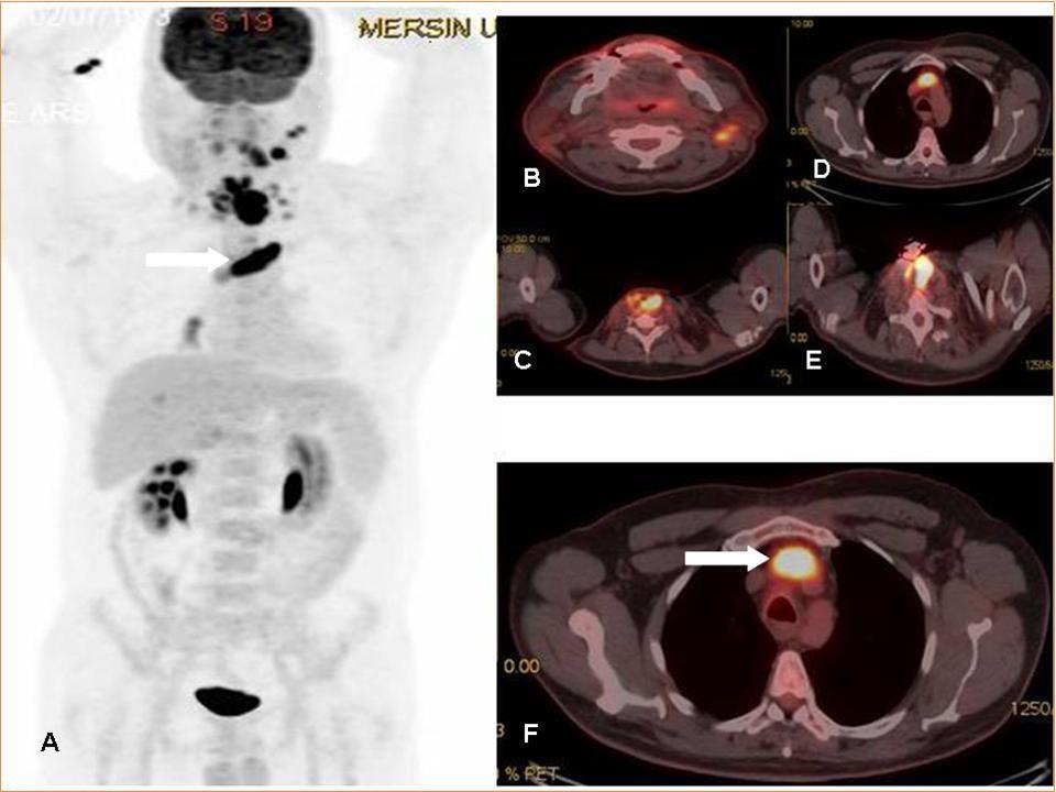
Download figure
References
- 1.Aurangabadkar H U, Palle L, Ali Z. (2013) Tumour thrombosis and patterns of fluorine-18 fluorodeoxyglucose uptake: a pictorial review. Nucl Med Commun. 34, 627-637.
- 2.Davidson T, Goitein O, Avigdor A. (2009) 18F-FDG-PET/CT for the diagnosis of tumor thrombosis. , Isr Med Assoc J; 11, 69-73.
- 3.Lai P, Bomanji J B, Mahmood S. (2007) Detection of tumour thrombus by 18FFDG-PET/CT imaging. , Eur J Cancer Prev; 16, 90-94.
- 4.Lee E Y, Khong P L. (2013) The value of 18F-FDG PET/contrast-enhanced CT in detection of tumor thrombus. , Clin Nucl Med; 38, 60-65.
- 5.Sharma P, Kumar R, Jeph S. (2011) 18F-FDG PET-CT in the diagnosis of tumor thrombus: can it be differentiated from benign thrombus?. , Nucl Med Commun; 32, 782-788.
- 6.Ravina M, Hess S, Chauhan M S, Jacob M J, Abass Alavi A. (2014) Tumor Thrombus. Ancillary Findings on FDG PET/CT in an Oncologic Population. , Clin Nucl Med; 39, 767-771.
- 7.Sonavane S N, Malhotra G, Asopa R, Upadhye T. (2015) Role of fluorine-18 fluorodeoxyglucose positron emission tomography in a case of renal cell carcinoma to differentiate tumor thrombus from bland thrombus. , Indian J Nucl Med.2015Oct-Dec; 30(4), 355-7.
- 8.Yang J, Zhen L, Zhuang H. (2013) Bone marrow metastases from alveolar rhabdomyosarcoma with impressive FDG PET/CT finding but less-revealing bone scintigraphy. , Clin Nucl Med.2013Dec; 38(12), 988-91.