Abstract
The postpartum period in camel are considered the most critical period for camel's health and for future fertility. The objective of the present study was to compare the clinical examination results and the concentration of hemoglobin, total protein, calcium and progesterone at different instances (1st, 3rd and 9th days) postpartum. Fifteen female camels during the postpartum period (1st, 3rd and 9th days) were included in the present study. Hematological, biochemical profiles and ultrasonographic examination were performed. The clinical examination results were normal and no evidence of clinical disease. The results of the hematological and biochemical findings were within the reference range obtained previously by our research team. The concentrations of hemoglobin, total protein, calcium and progesterone were measured at the 1st, 3rd and 9th days postpartum. Hemoglobin concentration differs significantly in the 3rd day (p=0.012) compared to the 1st day postpartum. Total protein showed significant increase in the 3rd day (p=0.003) compared to the 1st day postpartum. Calcium concentration showed significant increase in the 9th day (p=0.002) compared to the 1st and the 3rd days postpartum. Progesterone concentration showed significant decrease in the 9th day (p=0.004) compared to the 1st and the 3rd days postpartum. The ultrasonographic imaging of the uterus and ovaries showed normal uterine involution and no abnormal secretions in the uterus. It is concluded that, clinical examination, hematological and biochemical profiles are very important tools for monitoring female camels during postpartum period.
Author Contributions
Academic Editor: Roman Kireev, Institute of Biomedical Research Fundacion Biomedica Galicia Sur Hospital alvaro Cunqueiro - Bloque Tecnico - Planta 2 Estrada Clara Campoamor n 341 - 36312 Vigo, Spain
Checked for plagiarism: Yes
Review by: Single-blind
Copyright © 2017 Heba El-Zahar et al
 This is an open-access article distributed under the terms of the Creative Commons Attribution License, which permits unrestricted use, distribution, and reproduction in any medium, provided the original author and source are credited.
Competing interests
The authors have declared that no competing interests exist.
Citation:
Introduction
Transition period is an important part of the reproductive life of female camels due to its effect on future fertility1; it includes pre-partum and post-partum periods where many hematological and biochemical changes are observed. Most health disorders occur during the transition period resulting in severe economic losses2. In addition, the endocrine and metabolic changes associated with pregnancy, parturition and postpartum period are usually neglected during disease diagnosis3. The stressful condition during transition period and the decreased food intake with drop in the concentration of different blood constituents makes it very important in the animal’s life2, 4. Poor management during transition period might result in impairment of reproductive performance including delayed first service, long calving interval, relatively short breeding season and poor conception rate5, 6. Blood hematological and biochemical parameters are a well-known indicator for several metabolic processes in the body, which is usually differ between healthy and diseased camels and often vary due to age, sex, physiological conditions and environmental factors7, 8. The postpartum period has few studies and little attention during the last decades, the data describing the hematological and biochemical alterations are inadequate. However, the effect of the transition period on the hormonal changes in pregnancy, parturition and post-partum has been studied in llama3. The clinical monitoring of the animal’s health status during postpartum period parallel with hematological and biochemical analyses are proven to have an important impact on controlling postpartum diseases. Therefore, the objective of the present study is to compare the clinical examination results at different instances postpartum and the results of hemoglobin, total protein, calcium and progesterone at the 1st, 3rd and 9th days postpartum.
Materials and Methods
Camels
This study was performed on 15 female camels (Camelus dromedaries) during postpartum period. Camels are belonging to a private farm for camel breeding and management in Abu Dhabi, United Arab of Emirates, and the age was ranged from 6-9 years with an average body weight of 445kg. The study was proved by the animal wealth research unit of the Abu Dhabi Food Control Authority, United Arab of Emirates. The camels were apparently healthy on the basis of clinical examination and laboratory analysis and free from common infectious diseases as proved by general veterinary authorities. Ultrasound examination was done to confirm that these animals free from any reproductive disorders. The camels were group housed in an open yard daily with shelter all over the year and fed a properly formulated ration.
Clinical Examination
Clinical examination was performed for all camels included in the present study, restraining was made before examination. The clinical examination includes the following parameters; temperature, heart rate, respiration rate, mucous membrane, lymph node, capillary refill time, auscultation of the heart, lung and rumen9.
Blood Sampling
Two blood samples were drawn from the jugular vein at the 1st, 3rd and 9th days postpartum. Samples were collected in an EDTA tube for obtaining whole blood samples and on plain vacutainer for obtaining serum samples. Serum was harvested after centrifugation of the plain vacutainer at 1500 rpm for 10 minutes, and then stored at -20oC until analysis.
Hematological Analyses
Hematology profile were performed using Sysmex XT 2000i hematology analyzer (VLD-DPM-CBC-06, Mundelein, IL 60060, USA), for determination of erythrocytes, hematocrit, hemoglobin concentration, leucocytes, differential leucocytic count including neutrophils, lymphocytes, monocytes, eosinophils and basophils percentages. The hematological indices including MCV, MCH and MCHC were measured during analysis.
Biochemical Analyses
Biochemistry profile was performed using Beckman Coulter analyzers (VLD-DPM-CBC-02, Mundelein, IL 60060, USA). Total protein, albumin, creatinine, BUN, glucose, calcium, phosphorous and Iron (Fe) were measured in serum samples. The activity of serum enzymes including γGT, AST, ALT, creatine kinase and LDH were determined. Progesterone and estrogen concentration was determined in serum using ELISA kits (serum kits for e-procheck).
Ultrasonography
Ultrasonographic examination was performed for all camels included in the present study. The examination protocol was carried out in a daily basis until the 9th day postpartum then followed by examination twice weekly until the 21st day postpartum. Real-time, B-mode, ultrasound machine (Aloka, SSD 500, Tokyo, Japan) equipped with 5 MHz transrectal linear array transducer was used for examination. Uterus was palpated for consistency, contractility and examined ultrasonographically for lumen and content as well as vagina and cervix were examined for their normal passage into the uterus and for discharges.
Statistical Analysis
Statistical analysis was performed using SPSS Statistics® 24.0 (SPSS Inc., Chicago) and the results were expressed as mean values ± standard error (SE). Repeated measures ANOVA were used to compare multiple measures during postpartum period. The results were compared to those of non-pregnant camels. The level of significance was set at <0.05.
Results
In the present study, 15 female camels belonging to a private farm in UAE were used during the postpartum period and monitored in the 1st, 3rd and the 9th days postpartum. The clinical examination including temperature, heart and respiration rates, mucous membranes, lymph nodes, capillary refill time and auscultation of the heart, lung, rumen and intestine were normal and there is no evidence of clinical disease (Table 1).
Table 1. Mean values of clinical examination parameters in 15 female camels during the postpartum period (1st, 3rd and 9th days postpartum). The results are expressed as mean ± SE.| Parameters | Results |
|---|---|
| Temperature | 36.4±0.6 (35.5-37.2) (oC) |
| Heart rate | 44±1.2 (35-50) (beats/min.) |
| Respiration rate | 11 (7-16) (breaths/min.) |
| Mucous membrane | Normal rosy red |
| Capillary refill time | < 2 seconds |
| Lymph nodes | Different lymph nodes are of varying sizes, this was not indicative of disease |
| Heart auscultation | Normal systole diastole with normal rhythm |
| Lung auscultation | Normal vesicular sound |
| Rumen auscultation | Complete ruminal contraction is detected, 3±0.6 every 5 minutes |
The hematological and biochemical profile results are summarized in Table 2. The mean value of RBCs, hematocrit percent, erythrocyte indices MCV, MCH and MCHC, leucocytic count, neutrophils, lymphocytes, monocytes, eosinophils, basophils percents were within the reference range presented by our previous work7, there were no significant difference between the measurements of the three days postpartum (1st, 3rd and the 9th days postpartum). The concentration of albumin, glucose, iron, phosphorous, BUN, creatinine, creatine kinase, AST, ALT, LDH and γGT showed normal results compared to the results of previous work by Zaher et al.[7]; the results of estrogen showed significant decrease in the 3rd (18.06 pg/ml) and 9th (20.56 pg/ml) days postpartum compared to the 1st day (41.46 pg/ml), other biochemical parameters in Table 2 showed no significant difference.
Table 2. Selected hematological and biochemical parameters in 15 female camels during the postpartum period (1st, 3rd and 9th days postpartum). The results are expressed as mean ± SE.| 1 st day postpartum | 3 rd day postpartum | 9 th day postpartum | Sig. | |||||||
| Hematological profile | Erythrocytes (x106/ul3) | 9.75 | 9.96 | 10.12 | 0.082 | |||||
| Hematocrit (%) | 30.06 | 29.11 | 28.22 | 0.721 | ||||||
| MCV (fl) | 31.92 | 31.31 | 31.52 | 0.093 | ||||||
| MCH (pg) | 14.01 | 14.06 | 14.59 | 1.000 | ||||||
| MCHC (g/dL) | 47.52 | 47.01 | 46.85 | 0.931 | ||||||
| Leucocytes (x103/ul3) | 9.92 | 9.12 | 9.89 | 0.064 | ||||||
| Neutrophils (%) | 48.37 | 48.68 | 48.98 | 1.000 | ||||||
| Lymphocytes (%) | 37.66 | 40.12 | 38.43 | 1.000 | ||||||
| Monocytes (%) | 5.73 | 5.12 | 5.11 | 0.642 | ||||||
| Eosinophils (%) | 5.09 | 5.01 | 5.01 | 0.344 | ||||||
| Basophils (%) | 1.05 | 1.1 | 1.04 | 0.346 | ||||||
| Biochemical profile | Albumin (g/dL) | 3.63 | 3.54 | 3.57 | 0.327 | |||||
| Glucose (mg/dL) | 96.26 | 96.21 | 96 | 0.069 | ||||||
| Fe (ug/dL) | 110.78 | 112.32 | 114.3 | 0.627 | ||||||
| Phosphorous (mg/dL) | 5.68 | 5.67 | 5.82 | 0.248 | ||||||
| BUN (mg/dL) | 17.23 | 17.08 | 16.02 | 0.064 | ||||||
| Creatinine (mg/dL) | 0.91 | 0.92 | 0.99 | 0.082 | ||||||
| Creatine Kinase (U/L) | 86.33 | 86.41 | 85.69 | 1.000 | ||||||
| AST (U/L) | 92.51 | 90.55 | 91.99 | 1.000 | ||||||
| ALT (U/L) | 10.12 | 10.49 | 10.06 | 1.000 | ||||||
| LDH (U/L) | 587.97 | 662.18 | 616.59 | 0.095 | ||||||
| γGT (U/L) | 10.96 | 13.34 | 12.08 | 0.924 | ||||||
| Estrogen (pg/ml) | 41.46 | 18.06 | 20.56 | 0.046 | ||||||
The concentrations of hemoglobin, total protein, calcium and progesterone were measured at the 1st, 3rd and 9th days postpartum and the mean of the 3 measures were calculated. The mean values of the hemoglobin, total protein, calcium and progesterone in the 3 measures were within the reference ranges, although there were significant changes between the repeated measures (Table 3 and Table 4). Hemoglobin concentration significantly increased in the 3rd day (12.81 ± 0.426 g/dL) (p=0.012) compared to the 1st day postpartum (10.98 ± 0.176 g/dL) (Figure 1a). Total protein showed significant increase in the 3rd day (6.98 ± 0.201 g/dL) (p=0.003) compared to the 1st day postpartum (6.04 ± 0.112 g/dL) (Figure 1b). Calcium concentration showed significant increase in the 9th day (9.91 ± 0.209 mg/dL) (p=0.002) compared to the 1st (8.91 ± 0.084 mg/dL) and the 3rd days postpartum (9.6 ± 0.257 mg/dL) (Figure 1c). Progesterone concentration showed significant sharp decrease after parturition compared to the reference values of progesterone in normal conditions, in addition, there were marked significant decrease in the 9th day (0.25 ± 0.024 ng/mL) (p=0.004) compared to the 1st (0.45 ± 0.066 ng/mL) and the 3rd days postpartum (0.32 ± 0.039 ng/mL) (Figure 1d).
Table 3. Mean hemoglobin, total protein, calcium and progesterone concentration in 15 female camels in the 1st, 3rd and 9th days postpartum. The results are expressed as mean ± SE. * means significance at ≤0.05.| 1 st day postpartum | 3 rd day postpartum | 9 th day postpartum | Mean ± SE | Sig. | |
| Hemoglobin (g/dL) | 10.98 ± 0.176 | 12.81 ± 0.426 | 11.26 ± 0.26 | 11.69 ± 0.211* | 0.016 |
| Total protein (g/dL) | 6.04 ± 0.112 | 6.98 ± 0.201 | 6.22 ± 0.153 | 6.41 ± 0.109* | 0.006 |
| Calcium (mg/dL) | 8.91 ± 0.084 | 9.6 ± 0.257 | 9.91 ± 0.209 | 9.47 ± 0.128* | 0.001 |
| Progesterone (ng/mL) | 0.45 ± 0.066 | 0.32 ± 0.039 | 0.25 ± 0.024 | 0.34 ± 0.029* | 0.000 |
| Hemoglobin (g/ dL ) | Total protein (g/ dL ) | Calcium (mg/ dL ) | Progesterone (ng/mL) | ||
| 1st day PP | 3rd day PP | 0.012* | 0.003** | 0.074 | 1.000 |
| 9th day PP | 1.000 | 1.000 | 0.002** | 0.004** | |
| 3rd day PP | 9th day PP | 0.594 | 0.052 | 1.000 | 0.035* |
Figure 1.Illustrate the changes in the hemoglobin (a), total protein (b), calcium (c) and progesterone (d) during the 1st, 3rd and 9th days postpartum in female camels.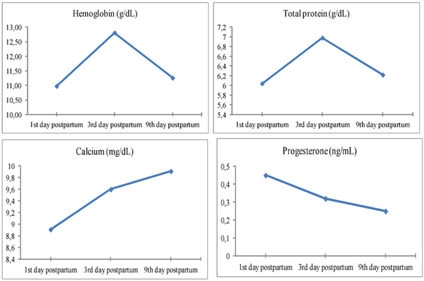
The ultrasonographic imaging of the uterus and ovaries was performed daily 9th day postpartum then followed by examination twice weekly until the 21st day postpartum. The ultrasound examination showed normal uterine involution and no abnormal secretions in the uterus (Figure 2).
Figure 2.Ultrasonographic image of ovaries (a) and uterine horn (b) at the 9th day postpartum in female camels, the image illustrate normal ovarian structure with no ovarian cysts and the uterus showed normal uterine involution and no abnormal discharges.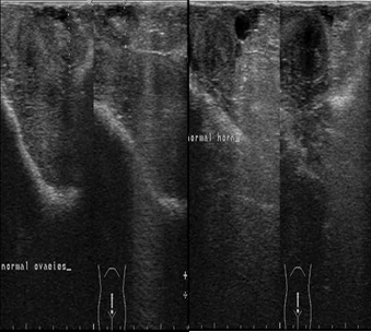
Discussion
The postpartum period is of highly clinical importance in female camels; although, it has gained little concern in camel practice. Good management during this period will secure healthy reproductive performance. The routine hematological and biochemical profiling during this period is of importance in predicting herds at risk of developing reproductive, metabolic and infectious diseases2. The present study was performed on 15 female camels during the postpartum period, the hematological and biochemical profiles were measured at the 1st, 3rd and 9th day postpartum. The clinical examination of female camels, including temperature, heart and respiration rates, mucous membranes, lymph nodes, capillary refill time and auscultation of the heart, lung, rumen and intestine, was given a great interest during the postpartum period to avoid most common problems occurred postpartum and to avoid deviations in the normal health status10. The hemoglobin concentration was an indication of anemia and blood status, where inappropriate feeding during the postpartum period might results in decreases hemoglobin levels. In the present study, the hemoglobin level was significantly increased during the 3rd day postpartum compared to the 1st and 9th days postpartum, however, it remains within the normal reference ranges. This can be taken as an indicator of the proper feeding regime and management during the transition period1. In sows, the hemoglobin values are decreased during pregnancy due to the metabolism of mother's hemoglobin in fetal circulation and the dilution of blood while during lactation it decreases11.
The total protein was considered an indicator of liver function and the immune status of female camels, in this study the results of total protein was concurred with the previous study obtained by our research team7, however a little variation between the 1st, 3rd and 9th days postpartum was observed and this might be attributed to immunoglobulin formation during the first few hours postpartum12. Also, the total protein concentration was increased in pregnant camels compared to non-pregnant which might be attributed to the immune status of the animal8. The serum calcium concentration is considered an important macromineral in female camels, it maintain normal body physiological condition, contraction of smooth and skeletal muscles and regulates nerve impulse transmission. The serum calcium concentration was lowered in pregnant than non-pregnant camels, which might be attributed to increased maternal demands in late pregnancy13. In the present study, the calcium concentration starts to increase gradually during the postpartum period which is considered an indicator of normal physiological condition after parturition and suggests normal uterine involution and reflects the well nourishment status of camels under this study14, 15.
The serum progesterone level decreased sharply after parturition during the 1st, 3rd and 9th days postpartum, which is considered physiological decrease due to the lysis of corpus luteum of pregnancy and the drop of placenta which is considered another source of progesterone to maintain pregnancy, in addition the lactation status significantly affects on serum progesterone level16. It was proved that the plasma concentration of progesterone was increased by the 5th days after mating and remained high throughout most of pregnancy, and it starts to decline by the 2nd weeks before parturition3, 17. In addition, the low level of progesterone detected in the 1st day postpartum concur with previous studies during postpartum females in camels18 and other species19. Similar changes in the hormonal levels were observed during pregnancy, parturition and postpartum in llama20, 21. The ultrasonographic examination was performed twice weekly during the postpartum period starting from the 3rd day postpartum until the 21st day postpartum, there was normal uterine involution and no abnormal secretions in the uterus, this was in agreement with previous studies by Abu-Seida22, Derar, Ali23, who found that the uterine involution was completed in female camels from 25th until 30th days postpartum.
Conclusion
In conclusion, clinical examination, hematological and biochemical profiles are very important tools for monitoring female camels during postpartum period. In addition, the level of progesterone and ultrasonographic examination of the uterus and ovaries give an indication about the normal uterine involution in parallel with normal hematologic and biochemical findings during the postpartum period.
References
- 1.Nazifi S, Ahmadi M R, Gheisari H R. (2008) Hematological changes of dairy cows in postpartum periodand early pregnancy. , Comp Clin Pathol; 17, 157-63.
- 2.Drackley J K. (1999) ADSA Foundation Scholar Award. Biology of dairy cows during the transition period: the final frontier?. , Journal of dairy science; 82, 2259-73.
- 3.Leon J B, Smith B B, Timm K I, LeCren G. (1990) Endocrine changes during pregnancy, parturition and the early post-partum period in the llama (Lama glama). , Journal of reproduction and fertility; 88, 503-11.
- 4.Abo-El maaty AM, El-Shahat K H. (2012) Hormonal and biochemical serum assay in relation to the estrous cycle and follicular growth in Arabian mare. Asian Pacific. , Journal of Reproduction; 1, 105-10.
- 6.Kaufmann B A. (2005) Reproductive performance of camels (Camelus dromedarius) under pastoral management and its influence on herd development. , Livestock Production Science; 92, 17-29.
- 7.Zaher H, El-Zahar H, Al Sharifi S, Shety T. (2017) Alterations in hematological and biochemical parameters affecting the reproductive performance in female camels. , International Journal of Veterinary Health Science & Research; 5, 155-60.
- 8.Ayoub M A, El-Khouly A A, Mohamed T M. (2003) Some hematological and biochemical parameters and steroid hormone levels in the one-humped camel during different physiological conditions. , Emir J Agric Sci; 15, 44-55.
- 9.Rosenberger G. (1990) Die Klinische Untersuchung des Rindes. 3rd ed. Berlin und Hamburg: Verlag Paul Parey;.
- 10.Tibary A, Anouassi A, Memon M A. (2001) Approach to diagnosis of infertility in camelids: Retrospective study in alpaca, lamas and camels. , Journal of camel practice and research; 8, 167-79.
- 11.Žvorc Z, Mrljak V, Sušić V, Pompe Gotal J. (2006) Haematological and biochemical parameters during pregnancy and lactation in sows. , Veterinarski arhiv; 76, 245-53.
- 12.Tharwat M, Oikawa S, Buczinski S. (2011) Ultrasonographic prediction of hepatic fat content in dairy cows during the transition period. , J Veterinar Sci Technol; 3, 111.
- 13.Saeed A, Khan I A, Hussein M M. (2009) Change in biochemical profile of pregnant camels (Camelus dromedarius) at term. , Comparative Clinical Pathology; 18, 139-43.
- 14.Kelanemer R, Antoine-Moussiaux N, Moula N, AAK Abu-Median, Hanzen C et al. (2015) Effect of nutrition on reproductive performance during the peri-partum period of femal camel (Camelus dromedarius) in Algeria. Journal of animal and veterinary advances; 14:. 192-6.
- 15.Muhammad B F, Aliyu D, Njidda A A, Madigawa I L. (2011) Some haematological, biochemical and hormonal profile of pregnant and non-pregnant she-camels (Camelus dromedarius) raised in a Sudan Savanna zone of Nigeria. , Journal of Camel Practice and Research(India);18
- 16.Kamoun M, Jemmali B. (2014) Serum progesterone level of camel (camelus dromedarius) according to the physiological status. , Journal of New Sciences; 3, 10-21.
- 17.Quzy I, Anwar S, Purohit G N. (2013) Hormonal management of ovarian activity in breeding camels two months ahead of the natural breeding season. , Camel: An International Journal of Veterinary Sciences; 1, 37-49.
- 18.Agarwal S P, Rai A K, Khanna N D. (1992) Hormonal studies in postpartum female camels and their neonates. , Theriogenology; 38, 735-47.
- 19.Henricks D M, Dickey J F, Hill J R, Johnston W E. (1972) Plasma oestrogen and progesterone levels after mating, during late pregnancy and postpartum in cows. , Endocrinology; 90, 1336-42.
- 20.Riveros J L, Schuler G, Bonacic C, Hoffmann B, Chaves M G et al. (2010) Ovarian follicular dynamics and hormonal secretory profiles in guanacos (Lama guanicoe). Animal reproduction science;. 119, 63-7.
- 21.Riveros J L, Urquieta B, Bonacic C, Hoffmann B, Bas F et al. (2009) Endocrine changes during pregnancy, parturition and post-partum in guanacos (Lama guanicoe). Animal reproduction science;. 116, 318-25.
Cited by (2)
- 1.Mahmood Nasir, Hameed Amjad, Hussain Tarique, Tutar Yusuf, 2020, Vitamin E and Selenium Treatment Alleviates Saline Environment-Induced Oxidative Stress through Enhanced Antioxidants and Growth Performance in Suckling Kids of Beetal Goats, Oxidative Medicine and Cellular Longevity, 2020(), 1, 10.1155/2020/4960507
- 2.الطيف طارق عبد السلام سالم, عامر منعم أبو القاسم, صابر سالم علي, امعزيق سالم ابوبكر, 2022, دراسة مقارنة لكرات الدم الحمراء وخصائصها بين النوق وحيرانها الذكور في ليبيا, Al-Mukhtar Journal of Sciences, 37(4), 412, 10.54172/mjsc.v37i4.922