Abstract
Background:
Furcation involvement is characterized by periodontal disease invading furcation regions of multi-rooted teeth. Treatment modalities are scaling and root planning and surgical management such as osteoplasty or ostectomy, odontoplasty, bicuspidization, root resection and hemisection. Periodontally compromised maxillary molars generally have poor prognosis because of inter radicular loss of attachment, and difficulty in access and treatment. Root resection is procedure by which one or more of the roots of a tooth are removed at level of furcation while leaving crown and the remaining roots in function.
Case:
A 58 year-old female patient applied to Periodontology clinic with complaints of sensibility and gingival recession in teeth number 16 and 26. Both teeth were completely exposed due to severe attachment loss in distobuccal root. There was also second degree mobility on the right molar and third degree mobility on the left molar teeth. Flapless root resection were planned after root canal therapy. After local anesthesia, distobucal roots were resected by high speed rotary motor with adequate irrigation at the level of the furcation roof. A small cavity was prepared and retrograded with glass ionomer cement. A platelet-rich fibrin membrane was obtained from patient’s blood and stitched to the distal surface of right first molar. Left first molar area was left uncovered. Recovery was followed on 2nd, 8th week and 6th month. When healing was compared between left and right sides, no mobility was observed and a slight redness and swelling was observed on the right side at 2nd week. At 8th week, there was no difference in clinical appearance. At 6th month, all complaints of the patient were gone and prognosis of the teeth was good.
Author Contributions
Academic Editor: Mario Perez Sayans, Associate Professor. University of Santiago de Compostela.
Checked for plagiarism: Yes
Review by: Single-blind
Copyright © 2017 Hatice Balci Yüce, et al
 This is an open-access article distributed under the terms of the Creative Commons Attribution License, which permits unrestricted use, distribution, and reproduction in any medium, provided the original author and source are credited.
Competing interests
The authors have declared that no competing interests exist.
Citation:
Introduction
Periodontal diseases are chronic infectious diseases which affects every part of society by causing unaesthetic appearance, tooth sensitivity, tooth mobility, alveolar bone loss and even tooth loss. Although primer etiologic factor is microbial dental plaque; 1 local factors such as root proximity, malalignment of the teeth, root grooves affect prognosis of the disease especially in multi-rooted teeth. Non-surgical periodontal treatment has been reported to be successful in single rooted teeth; but multi-rooted teeth which have local predisposing factors might require additional precautions 2. Regenerative therapies also provide limited improvement unless these factors are not eliminated 3. Studies have shown that tooth loss during long-term supportive periodontal treatment is 31-57% in molar teeth and 5-7% in single-rooted teeth 4, 5. Furthermore, Buduneli et al. performed non-surgical periodontal treatment in 40 single-rooted teeth of 18 patients with chronic periodontitis and recorded changes after 1 year. According to the results of the study, the papillary hemorrhage index decreased from 3.63 to 1.67, the mean pocket depth decreased from 7.89 mm to 4.84 mm and the mean attachment gain was 2.12 mm 6. Therefore, periodontal treatment in molar teeth is usually more complicated than anterior teeth.
Periodontal disease can cause alveolar bone resorption and connective tissue attachment loss by influencing the furcation of multi-rooted teeth. After involvement of furcation, the lesion becomes a furcation defect 7, 8. Involvement of furcation makes it difficult to obtain successful treatment results due to the anatomy of this region, inability to perform effective scaling and root planning (SRP) and inadequate plaque control 9, 10, 11, 12. Root anatomy (concavities, grooves), furcation and deep sites are the limiting factors for an effective SRP procedure 13. Single-rooted teeth are the most benefited teeth from SRP.Maxillary molar teeth have mesial and distal furcation insertions, and inflammatory lesions may result in loss of the interdental bone of the adjacent tooth. In extreme cases, when the lesion progresses, and whole root becomes completely exposed; the prognosis of one root is worse than the relevant tooth. SRP would be ineffective and alternative treatment modalities should be considered. In such times, root resection is one of the treatment choices.
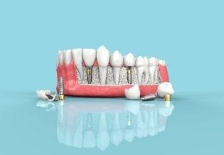
Root resection is the procedure in which one or more roots of a tooth are separated at the level of the furcation, leaving the remaining roots in the function. Farrar has identified this root resection procedure to treat Class II and Class III furcation lesions in molar teeth 14. This process creates cleanable, accessible areas for the clinician and the patient.
Case
A 58-year-old female patient applied to the Periodontology clinic with the complaints of sensitivity and gingival recession in both maxillary first molar teeth. Patient had no systemic diseases, no smoking habit or any other condition. Both second molar teeth of the patient were missing and other than first molar teeth, there was no decay or gingival inflammation. Canine, premolars and first molar were in full contact with mandibular teeth. Patient had no bruxism and temporomandibular joint problem. The only dental problem of the patient was maxillary first molar teeth. Distobuccal roots of both teeth were completely exposed due to severe attachment loss. There was a small portion of soft tissue around right molar’s distobuccal root and no soft tissue was left around left first molar’s distobuccal root. Plaque and gingival index of the teeth were score ‘1’ in both teeth. Probing depth was 2 mm in right maxillary molar teeth and 2.5 mm in left maxillary molar teeth. Other roots of right molar had no attachment loss while the distal aspect of left molar’s mesiobuccal root had severe attachment loss. Left molar tooth had grade III mobility and right molar tooth had grade II mobility. Both of teeth were tender on percussion and had no decay. Roots were completely exposed and a flapless root resection was performed after root canal therapy. There was no need for local anesthesia for right molar but an infiltrative local anesthesia was performed on soft tissues around left molar’s root. Distobuccal roots were resected from the furcation roof by high speed rotary motor with adequate irrigation. A small cavity was prepared for the exposed root canal and retrograded with glass ionomer cement. For right molar, Platelet Rich Fibrin (PRF) was placed and sutured with 4-0 silk suture (Ethicon, Somerville, New Jersey, USA) to cover the soft tissues after resection but left molar was left uncovered. In order to obtain PRF, 10 mL venous blood was driven and centrifuged in an empty glass tube (2800 rpm, 12 min.) PRF was sutured with four interrupted sutures and a horizontal mattress suture from the corners. CHX mouth rinse and analgesics were prescribed and oral hygiene maintenance instructions and post-surgical instructions were given. Sutures were removed after two weeks. Mobility in both teeth decreased to grade I. Recovery was followed on 2nd week, 6th week and 6th month. Sutures were removed at 2nd week. At 6th month evaluation, all complaints of the patient were gone.
Discussion
Root resection is a treatment option for multi-rooted teeth with endodontic, periodontal or prosthetic reasons. Patient's oral hygiene status, caries index and medical condition should be considered in order to obtain a successful result. The success rate of the procedure depends on bone support of the other roots, occlusion, patient’s oral hygiene, and effective root-canal treatment 15, 16.
The prognosis of root resection has been well-documented in previous studies 8, 17, 18, 19. The 4-year survival rate of maxillary molars after root amputation is reported to be 93% 20 and long-term survival of teeth after root amputation ranges from 87% to 95% 15. In present case, there were three treatment options. 1; follow up after non-surgical periodontal treatment and endodontic treatment without surgical intervention; 2; root resection after non-surgical periodontal treatment and endodontic treatment lastly, 3; extraction of teeth. In present case, distobuccal roots of maxillary first molar teeth were excised. Remaining roots provided adequate support for teeth to survive and there was no need for tooth extraction. Root resection was successful in a short term period such as six months. In present case, the patient was 58 years old and there was no sign of any other periodontal disease in the mouth. For this reason, there is no plausible explanation for the etiology of the furcation lesions in this patient. The aforementioned local anatomical factors or previous traumatic extraction of second molar may have set the ground for the loss of attachment in distobucal roots. The severity of furcation lesion was lower in the right side and a better improvement was observed. In addition, PRF was sewn to the wound area after the resection of the right upper first molar's distobuccal root and PRF could have improved healing process as shown by other studies in the literature. Left molar tooth had also bone loss around mesiobuccal root and therefore there is a risk of further problems in this area.
Conclusion
Root resection is usually performed for advanced furcation lesions which generally indicate severe periodontal disease. However, in present case, we performed root resection in a patient who had no other bone loss except maxillary right and left first molar teeth. As a conclusion, resecting the most affected root of a severe maxillary furcation defect improved clinical condition of the tooth and the prognosis also improved. Root resection can be performed as a last treatment option instead of extracting tooth in such cases.
References
- 1.Tezal M. (2006) Supragingival plaque may modify the effects of subgingival bacteria on attachment loss. Journal of periodontology. 77(5), 808-813.
- 2.Nieminen A. (1995) Prognostic criteria for the efficiency of non‐surgical periodontal therapy in advanced periodontitis. Journal of clinical periodontology. 22(2), 153-161.
- 3.Evans G. (1995) Frequency of furcation closure with regenerative periodontal therapy. in The Journal of the Western Society of Periodontology/Periodontal abstracts. 44-4.
- 4.Hirschfeld L, Wasserman B. (1978) A long-term survey of tooth loss in 600 treated periodontal patients. Journal of periodontology. 49(5), 225-237.
- 5.McFall Jr WT. (1982) Tooth loss in 100 treated patients with periodontal disease: a long-term study. , Journal of Periodontology 53(9), 539-549.
- 6.Buduneli E. (2001) Comparative clinical and microbiological effects of subgingival metronidazole application in adult periodontitis; 12-months results. , Journal of the International Academy of Periodontology 3(4), 81-86.
- 7.Ward C. (1999) Furcation depth and interroot separation dimensions for 5 different tooth types. , International Journal of Periodontics & Restorative 19(3).
- 8.Walter C, Weiger R, N U Zitzmann. (2011) Periodontal surgery in furcation-involved maxillary molars revisited—an introduction of guidelines for comprehensive treatment. Clinical oral investigations. 15(1), 9-20.
- 9.Auplish G. (2000) Diamond‐coated sonic tips are more efficient for open debridement of molar furcations. Journal of clinical periodontology. 27(5), 302-307.
- 10.Svärdström G, J L Wennström. (2000) Periodontal treatment decisions for molars: an analysis of influencing factors and long-term outcome. Journal of periodontology. 71(4), 579-585.
- 11.Lindhe J. (1984) Long‐term effect of surgical/non‐surgical treatment of periodontal disease. , Journal of clinical periodontology 11(7), 448-458.
- 12.Kalkwarf K L, W B Kaldahl, K D Patil. (1988) Evaluation of furcation region response to periodontal therapy. , Journal of periodontology 59(12), 794-804.
- 13.Machtei E E, Ben-Yehouda A. (1994) The effect of post-surgical flap placement on probing depth and attachment level: a 2-year longitudinal study. , Journal of periodontology 65(9), 855-858.
- 14.J N Farrar.Radical and heroic treatment of alveolar abscess by amputation of roots of teeth, with description and application of the cantalever crown. 1884: Philadelphia:: SS White Dental Manufacturing Company.
- 15.Gupta A, Kabra P.Resective Procedure in the Management of Maxillary Molar with Horizontal/Oblique Root Fractures: A Case Report. IOSR Journal of Dental and Medical Sciences (IOSR-JDMS) 1(15), 66-69.
- 17.Minsk L, Polson A. (1995) The role of root resection in the age of dental implants. Compendium of continuing education in dentistry. , Jamesburg, NJ: 27(7), 384.
- 18.Carnevale G, Pontoriero R, Febo G D. (1998) Long‐term effects of root‐resective therapy in furcation‐involved molars. , Journal of Clinical Periodontology 25(3), 209-214.